SciELO - Scientific Electronic Library Online

 
vol.30 número1Aterosclerosis Carotidea: Una causa frecuente pero prevenible de ictus isquémico.Encefalopatía Hipóxico-Isquémica Por Anfetaminas índice de autoresíndice de materiabúsqueda de artículos
Home Pagelista alfabética de revistas  

Servicios Personalizados

Revista

Articulo

Indicadores

Links relacionados

  • No hay articulos similaresSimilares en SciELO

Compartir


Revista Ecuatoriana de Neurología

versión On-line ISSN 2631-2581versión impresa ISSN 1019-8113

Rev Ecuat Neurol vol.30 no.1 Guayaquil abr./jul. 2021

https://doi.org/10.46997/revecuatneurol30100185 

Imágenes en Neurología

Temporal Squama Pneumatization: An Under-Recognized Limitation Of Transcranial Doppler.

Pneumatización De Las Escamas Temporales: Una Limitación Poco Reconocida Del Doppler Transcraneal.

Oscar H. Del-Brutto1 

Juan A. Nader2 

1Universidad de Especialidades Espíritu Santo, School of Medicine. Guayaquil, Ecuador. <oscardelbrutto@hotmail.com>

2Hospital Médica Sur, Department of Neurology. Mexico City, Mexico.


Key Words: Transcranial Doppler; Acoustic windows; Poor insonation; Temporal squama pneumatization.

Palabras clave: Doppler transcraneal; ventanas acústicas; pobre insonación; escama temporal; pneumatización.

Images in Neurology

Transcranial Doppler (TCD) is a non-invasive procedure that is increasingly used for diagnostic and prognostic purposes in patients with an acute stroke (1). In addition, TCD enhances the effect of thrombolysis by exposing thrombi surfaces to circulating rTPA(2). However, insonation problems through acoustic windows limit the diagnostic efficiency of TCD. Accurate prediction of individuals who are more prone to present such problems may save valuable time for decision making on thrombolysis. Increased thickness of temporal bones and the presence of exuberant cancellous bone (diploe) account for the high rate of TCD failures among women, older individuals and people of different ethnic groups (3)(4). In addition, air may also interfere with proper assessment of signal flows from intracranial arteries. Aberrant pneumatization of temporal squamas were recognized as responsible for TCD failures in only one study(5), but no further attention has been given to this condition, which is thought to be more common in persons with recurrent ear infections, nasal deflections or in those exposed to frequent high altitude travel or Valsalva maneuvers(6). Routine evaluation of bone window on CT scans may help to visualize aberrant pneumatization of the temporal squama and to identify individuals who are not candidates for TCD. (Figure 1)

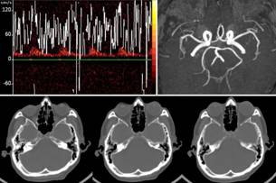
Figure 1 Upper panel: Poor trans-temporal windows insonation of the left middle cerebral artery (right MCA was not visualized) during TCD in a 70-year-old man with patent MCAs as demonstrated by magnetic resonance angiography. Lower panel: CT with bone window shows extensive pneumatization of temporal squamas, which extended more than 1cm anteriorly to the insertion of the pinna. 

References

Purkayastha S, Sorond F. Transcranial Doppler ultrasound: technique and application. Semin Neurol 2012; 32: 411-420. https://doi.org/10.1055/s-0032-1331812 [ Links ]

Alexandrov AV. Ultrasound enhanced thrombolysis for stroke. Int J Stroke 2006; 1: 26-29. http://doi.org/10.1111/j.1747-4949.2005.00012.x [ Links ]

Kwon JH, Kim JS, Kang DW, Bae KS, Kwon SU. The thickness and texture of temporal bone in brain CT predict acoustic window failure of transcranial Doppler. J Neuroimaging 2006; 16: 347-352. http://doi.org/10.1111/j.1552-6569.2006.00064.x [ Links ]

Del Brutto OH, Mera RM, Andrade ML, Espinosa V, Castillo PR, Zambrano M, et al. Temporal bone thickness and texture are major determinants of the high rate of insonation failures of transcraneal Doppler in Amerindians (the Atahualpa Project). J Clin Ultrasound 2016; 44: 55-60. http://doi.org/10.1002/jcu.22284 [ Links ]

Jarquin-Valdivia AA, McCartney J, Palestrant D, Johnston SC, Gress D. The thickness of the temporal squama and its implication for transcranial sonography. J Neuroimaging 2004; 14: 139-142. https://doi.org/10.1111/j.1552-6569.2004.tb00230.x [ Links ]

Rebol J, Munda A, Tos M. Hyperpneumatization of the temporal, occipital and parietal bones. Eur Arch Otorhinolaryngol 2004; 261: 445-448. https://doi.org/10.1007/s00405-003-0716-6 [ Links ]

Funding This study was partially supported by University Espíritu Santo - Ecuador, Guayaquil - Ecuador, and Medica Sur Foundation Clínica, Mexico City, Mexico.

Received: January 04, 2021; Accepted: February 04, 2021

Conflict of interest

Nothing to disclose.

Creative Commons License This is an open-access article distributed under the terms of the Creative Commons Attribution License