SciELO - Scientific Electronic Library Online

 
vol.26 número3Apnea obstructiva del sueño y neumatizaciòn aberrante de huesos del cráneo.Funciones Ejecutivas En Niños Preescolares Con Y Sin Trastorno Del Lenguaje índice de autoresíndice de materiabúsqueda de artículos
Home Pagelista alfabética de revistas  

Servicios Personalizados

Revista

Articulo

Indicadores

Links relacionados

  • No hay articulos similaresSimilares en SciELO

Compartir


Revista Ecuatoriana de Neurología

versión On-line ISSN 2631-2581versión impresa ISSN 1019-8113

Rev Ecuat Neurol vol.26 no.3 Guayaquil sep./dic. 2017


Artículo Original

Cerebral pulsatility index in older adults with silent lacunar strokes (The Atahualpa Project).

Índice de pulsatilidad cerebral en adultos mayores con infarto lacunar silente (Proyecto Atahualpa).

Oscar Del Brutto MD1 

Robertino Mera MD2 

Pablo Castillo MD3 

María de la Luz Andrade MD4 

Mauricio Zambrano BS5 

Juan Nader MD4 

1Universidad de Especialidades Espíritu Santo, School of Medicine, Guayaquil, Ecuador, oscardelbrutto@hotmail.com

2Vanderbilt University, Gastroenterology Department, Nashville, TN

3Mayo Clinic College of Medicine, Sleep Disorders Center, Jacksonville, FL

4Hospital Medica Sur, Department of Neurology, Mexico City, Mexico

5The Atahualpa Project, Community Center, Atahualpa, Ecuador


ABSTRACT

Background:

Diagnosis of silent lacunar infarcts is complicated in remote rural areas where MRI is not available. Hospital series have suggested an association between the pulsatility index of intracranial arteries - as assessed by transcranial Doppler - and some neuroimaging signatures of cerebral small vessel disease. We aimed to assess the reliability of cerebral pulsatility indices to identify candidates for MRI screening in population-based studies assessing prevalence of silent lacunar infarctions.

Methods:

A random sample of stroke-free Atahualpa residents aged ≥60 years investigated with MRI underwent transcranial Doppler for calculating the pulsatility index (PI) of the middle cerebral artery (MCA). For each person, mean PI was obtained by averaging both MCAs. Using conditional logistic regression for matched pairs data, we evaluated whether the pulsatility index of both MCAs correlate with silent lacunar infarcts.

Results:

Silent lacunar infarcts were noticed in 28 (12%) of 234 scanned persons. Six of them were excluded due to poor insonation through transtemporal windows. The remaining 22 participants were considered case-patients and were matched 1:1 with individuals free of infarcts (controls). Moderate-to-severe white matter hyperintensities were noticed in 12 (55%) case-patients and 7 (32%) controls (p=0.228). The mean MCA PI value in the 44 participants was 1.15 ± 0.21, with no difference found across case-patients and controls, after adjustment for white matter hyperintensities (β coefficient: 3.361, 95% C.I.: -0.693 to 7.417, p=0.104).

Conclusions:

Cerebral PI should not be used to identify candidates for MRI screening in population-based studies assessing the burden of silent lacunar infarcts.

Keywords: Lacunar infarction; transcranial doppler; pulsatility index

RESUMEN

Antecedentes:

El diagnóstico de infartos lacunares silentes es complicado en áreas rurales donde no se dispone de IRM. Series hospitalarias han sugerido una asociación entre el índice de pulsatilidad (IP) de las arterias intracraneales -evaluadas mediante Doppler transcraneal- y algunas de las imágenes sugestivas de enfermedad cerebral de pequeños vasos. Nuestro objetivo fue evaluar la confiabilidad del IP para identificar candidatos para la práctica de IRM en estudios poblacionales que evalúen prevalencia de infartos lacunares.

Métodos:

Una muestra aleatoria de residentes de Atahualpa sin evidencia de ictus, con edad ≥60 años e investigados con IRM, fueron sometidos a Doppler transcraneal, para calcular el IP de las arterias cerebrales medias. El IP medio se obtuvo promediando el IP de ambas arterias cerebrales medias. Usando regresión logística condicional, se evaluó si el IP de las arterias cerebrales medias se correlacionó con la presencia de infartos lacunares.

Resultados:

Se detectaron infartos lacunares silentes en 28 (12%) de 234 personas exploradas. Seis de ellos fueron excluidos debido a mala insonancia a través de ventanas transtemporales. Los 22 participantes restantes se consideraron casos y se emparejaron 1: 1 con individuos libres de infartos (controles). Se observaron hiperintensidades de sustancia blanca de moderada a grave en 12 (55%) pacientes y 7 (32%) controles (p = 0.228). El valor medio de IP en los 44 participantes fue de 1,15 ± 0,21, sin diferencias entre pacientes y controles, después del ajuste para hiperintensidades de sustancia blanca (coeficiente β: 3,361, 95% C.I.: -0,693 a 7,417, p = 0,104).

Conclusiones:

El IP cerebral no se debe utilizar para identificar candidatos para el cribado de IRM en estudios poblacionales que evalúen la presencia de infartos lacunares silentes.

Palabras clave: Infarto lacunar; doppler transcraneal; índice de pulsatilidad

INTRODUCTION

Stroke burden is increasing in rural areas of Latin America1. Identification of mechanisms underlying stroke may be useful for the implementation of interventions in apparently healthy individuals before cerebrovascular events occur. A recent study conducted in older adults living in rural Ecuador showed that two thirds of these persons have MRI evidence of silent cerebral small vessel disease (SVD), emphasizing the important of this condition as a major (and hidden) public health problem2.

Diagnosis of SVD requires the use of MRI, which is not readily available in remote rural settings. Efforts should be directed to find portable screening diagnostic tools that help to identify candidates for MRI screening. Transcranial Doppler (TCD) examination has been proposed as an inexpensive alternative. The pulsatility index (PI) - as calculated by transcranial Doppler (TCD) - has been considered a proxy of cerebral SVD as it may reflect distal cerebrovascular resistance. However, the literature on this subject is inconclusive3,4,5,6,7. We conducted a population-based and case-control nested study in an Ecuadorian rural village to evaluate whether the PI correlates with the presence of silent lacunar infarcts (a recognized neuroimaging signature of cerebral SVD).

METHODS

The IRB of Hospital-Clínica Kennedy, Guayaquil - Ecuador (FWA 00006867) approved the protocol and the written informed consent. Out of 297 stroke-free Atahualpa residents aged ≥60 years identified during a door-to-door survey, 234 underwent MRI/MRA. Exams were performed with a Philips Intera 1.5T MRI machine (Philips Medical Systems, the Netherlands), using previously described protocols2,8. Primary interest focused on the presence of lacunar infarcts, defined as fluid-filled cavities measuring 3-15mm located in the territory of a perforating arteriole9. White matter hyperintensities (WMH) of presumed vascular origin, defined as lesions appearing hyperintense on T2-weighted images that remained bright on FLAIR (without cavitation), were noticed and graded according to the modified Fazekas scale10. On MRA, the presence of ≥50 stenosis of one or both MCAs excluded the person from the analysis. In addition, individuals with an overt stroke were identified by certified neurologists and were not included in this study.

Participants with silent lacunar infarcts (case-patients) were matched 1:1 by age and sex with individuals who had a normal MRI (controls). Statistical significance was tested by the conditional logistic regression for matched pairs data, which is the preferred analytical tool for matched case-control studies

RESULTS

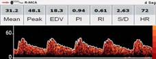
Silent lacunar infarcts were noticed in 28 (12%) of 234 scanned persons. They underwent TCD with the use of a SONARA portable system (VIASYS Healthcare, Inc. Madison, WI, USA) and a 2-MHz probe, following a well-known power motion mode Doppler/spectral TCD protocol11. Insonation problems precluded assessment of flow signals from MCAs in six of these individuals, which were excluded. In the remaining 22 participants, the pulsatility index (PI) of MCAs were calculated using the Gosling equation (peak systolic velocity-end-diastolic velocity/mean flow velocity). For each person, mean PI was obtained by averaging both MCAs.

As case-patients and controls were properly matched, there no difference in the mean age (73.8 ± 6.6 versus 73.2 ± 6.7, p=0.766) or in the percentage of men (55%) across groups. Moderate-to-severe WMH was noticed in 12 (55%) case-patients and 7 (32%) controls, with no differences across groups (p=0.228, McNemar’s test). The mean MCA PI value in the 44 participants was 1.15 ± 0.21, with no difference found across case-patients and controls when significance was tested by the conditional logistic regression for matched pairs data, after adjustment for WMH (β coefficient: 3.361, 95% C.I.: -0.693 to 7.417, p=0.104). Indeed, we noticed a flattening systolic peak in TCD in several cases with silent lacunar infarcts, with the formation of a particular type of wave, which makes a plateau instead of a high PI (Figure 1).

Figure 1 Flattening systolic peak of the middle cerebral artery in transcranial Doppler of a patient with a silent ipsilateral lacunar infarct. Notice the formation of a particular type of wave, which makes a plateau instead of a high pulsatility index. 

DISCUSSION

This case-control study, conducted in community-dwelling older adults living in a remote rural setting, shows lack of association between MCA PI and silent lacunar infarcts, suggesting that a high PI might be unrelated to SVD. A high PI may not only reflect distal cerebrovascular resistance (and thus, SVD) but may also occur as the result of large artery stiffness or other hemodynamic factors.12) Because of its complex nature, PI is not useful to assess prevalence of silent lacunar infarctions and should not be used alone as a proxy for silent lacunar infarcts.

The small sample size is a limitation of this study. However, the fact that we included all persons with silent lacunar infarcts found in the community, together with the case-control design argue for the strength of our findings. Further studies are needed before proposing that a high PI should be used to guide the practice of MRI at the community level for estimating the prevalence of SVD.

REFERENCES

Lavados PM, Hennis A, Fernandes JG, et al. Stroke epidemiology, prevention, and management strategies at a regional level: Latin America and the Caribbean. Lancet Neurol 2007; 6: 362-372. [ Links ]

Del Brutto O.H, Mera R.M, Del Brutto V.J, Zambrano M, Lama J. White matter hyperintensities of presumed vascular origin: a population-based Study in rural Ecuador (The Atahualpa Project). Int J Stroke 2015; 10: 372-375. [ Links ]

Kidwell CS, El-Saden S, Livshits Z, Martin NA, Glenn TC, Saver JL: Transcranial Doppler pulsatility indices as a measure of diffuse small-vessel disease. J Neuroimaging 2001; 11: 229-235. [ Links ]

Mok V, Ding D, Fu J, et al: Transcranial Doppler ultrasound for screening cerebral small vessel disease. A community study. Stroke 2012; 43: 2791-2793. [ Links ]

Heliopoulos I, Artemis D, Vadikolias K, Tripsianis G, Piperidou C, Tsivgoulis G: Association of ultrasonographic parameters with subclinical white-matter hyperintensities in hypertensive patients. Cardiovasc Psychiatry Neurol 2012; 2012: 616572. [ Links ]

Han SW, Song TJ, Bushnell CD, et al: Cilostazol decreases cerebral arterial pulsatility in patients with mild white matter hyperintensities: subgroup analysis from the effect of cilostazol in acute lacunar infarction based on pulsatility index of transcranial Doppler (ECLIPse) study. Cerebrovasc Dis 2014; 38: 197-203. [ Links ]

Del Brutto OH, Mera RM, Andrade MdeL, Castillo PR, Zambrano M, Nader JA: Disappointing reliability of pulsatility indices to identify candidates for magnetic resonance imaging screening in population-based studies assessing prevalence of cerebral small vessel disease. J Neurosci Rural Pract 2015; 6: 336-338. [ Links ]

Del Brutto OH, Mera RM, Zambrano M, Lama J: Incompleteness of the circle of Willis correlates poorly with imaging evidence of small vessel disease. A population-based study in rural Ecuador (the Atahualpa Project). J Stroke Cerebrovasc Dis 2015; 24: 73-77. [ Links ]

Wardlaw JM, Smith EE, Biessels GJ, et al. Neuroimaging standards for research into small vessel disease and its contribution to ageing and neurodegeneration. Lancet Neurol 2013; 12: 822-838. [ Links ]

Pantoni L, Basile AM, Pracucci G, et al: Impact of age-related cerebral white matter changes on the transition to disability: the LADIS study: rationale, design and methodology. Neuroepidemiology 2005; 24: 51-62. [ Links ]

Alexandrov AV, Demchuk AM, Burgin WS: Insonation method and diagnostic flow signatures for transcranial power motion (M-mode) Doppler. J Neuroimaging 2002; 12: 236-244. [ Links ]

Webb AJ, Simoni M, Mazzucco S, Kuker W, Schulz U, Rothwell PM. Increased cerebral arterial pulsatility in patients with leukoaraiosis: arterial stiffness enhances transmission of aortic pulsatility. Stroke 2012; 43: 2631-2636. [ Links ]

Received: September 05, 2017; Accepted: December 05, 2017

Conflict of interest Nothing to disclose.

Funding Study supported by Universidad Espíritu Santo - Ecuador, Guayaquil - Ecuador, and Médica Sur Fundación Clínica, Mexico City, Mexico.

Creative Commons License This is an open-access article distributed under the terms of the Creative Commons Attribution License