


 Inglés (pdf)
 Articulo en XML
 Referencias del artículo
 Enviar articulo por email
 Citado por SciELO
  Similares en
SciELO
Similares en
SciELO 
 Permalink
IMAGES IN NEUROLOGY
The external capsule is a band of longitudinal fibers (white matter) limited by two deep gray matter structures, the putamen medially and the claustrum laterally (Figure 1). This structure is mainly composed of axons that connect different areas of the cerebral cortex with the tegmentum (corticotegmental fibers). Ischemic strokes confined to the external capsule are extremely rare, representing 0.3% of patients enrolled in a large hospital-based ischemic stroke registry (1). External capsule infarcts may be related to different pathogenic mechanisms, including large artery disease, cardiogenic brain embolism, sporadic cerebral small vessel disease, or to a combination of them. In addition, external capsule infarcts have been typically reported in a hereditary form of cerebral small vessel disease known as CADASIL (cerebral autosomal dominant arteriopathy with subcortical infarcts and leukoencephalopathy) (2).
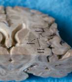
Figure 1 Anatomical specimen at the level of the basal ganglia showing the external capsule and its two boundaries, the putamen and the claustrum (arrows).
External capsule infarcts related to large artery atherosclerotic disease are not common. Indeed, only three out of nine patients with these infarcts had significant stenosis of the internal carotid artery as the most likely cause (1). In none of these patients there was isolated occlusion or stenosis of the middle cerebral artery.
While arterial supply of the external capsule mostly comes from lateral lenticulostriate branches of the middle cerebral artery, this structure is mostly a borderzone area and its ischemic damage depends, in part, of collateral blood supply. The case reported here is a clear example of an isolated external capsule infarct related to occlusion of the M1 segment of the middle cerebral artery (Figure 2).

Figure 2 A 74-year-old man with history of arterial hypertension presented with right facio-brachial paresis of sudden onset associated with ipsilateral sensory deficit. Magnetic resonance angiography of intracranial vessels showed occlusion of the left middle cerebral artery (left panel), and T1-weighted inversion-recovery sagittal sequence showed an infarct located at the left external capsule (right panel).

