


 Inglés (pdf)
 Articulo en XML
 Referencias del artículo
 Enviar articulo por email
 Citado por SciELO
  Similares en
SciELO
Similares en
SciELO 
 Permalink
Introduction
PD is a chronic degenerative disease with severe loss of DAergic cells from the nigrostriatal pathway in the brain. The World Health Organization estimates that over 5.8 million people in the world suffer from PD. In Mexico, the estimated incidence of PD is 14.9/100,000 in 2023 (1).
One of the available options to treat PD is pharmacological therapy. The drugs used for this purpose aim to normalize the motor function of the patients and try to avoid the appearance of adverse effects. Although DA is an endogenous neurotransmitter of the CNS and its severe loss causes the motor symptoms observable in the PD, it cannot be administered systemically to restore the DAergic neurotransmission and the normal motor functions due to its inability to cross the blood-brain barrier (BBB). For this reason, other drugs that can cross the BBB, such as L-DOPA, a precursor of DA synthesis, or DA agonists are used. L-DOPA is the most effective drug in reducing the symptoms of PD. However, after prolonged use, almost 50% of patients develop significant motor fluctuations and dyskinesias (2). The medical costs of treating patients with PD increase in direct proportion to the onset of these complications. In addition, antiparkinsonian drugs frequently have poor or discontinuous availability (3).
Recent studies (4)(5)(6) have suggested the possibility of using nanomaterials from inorganic matrices like silicon dioxide (SiO2) as a biocompatible excipient for the controlled release and delivery of various organic molecules to their target sites. When the matrices meet in contact with the tissue, the molecules contained in the excipient are released through the pores on the surface of the material in a sustained form, presenting an interesting alternative in the field of drug delivery (5). In the study of López et al (4). in hemiparkinsonian rats, it was found that the implant in the striatum of dioxide-silica with DA (SiO2-DA), significantly reduced the number of turns induced by systemic administration of apomorphine in relation to the behavior before the lesion. However, the symptomatology of PD is complex. Patients with PD show motor symptoms such as tremors at rest, decrease and delay in the initiation of movements (akinesia), slowness in movements (bradykinesia), rigidity, and postural instability (1). The reduction of the circling behavior is only one parameter of the beneficial effect on motor activity of treatment in hemiparkinsonian rats. A symptom that presents in human patients of PD and chronic models of parkinsonism in animals is postural instability. The tests used to evaluate the beneficial effect of the treatments against PD in animal models have clinical relevance. The objective of the present study is to assess the effectiveness of intrastriatal implants of SiO2-DA to recover postural stability in animals with massive loss of DAergic neurons of the SNpc with 6-OHDA. This design also allows for the assessment of the instauration of side effects such as motor oscillations or abnormal responses such as dyskinesia caused by the prolonged systemic administration of antiparkinsonian drugs such as L-DOPA. It also allows monitoring of the development of complications due to the surgical procedure itself. The findings obtained in this study will provide insight into the viability of assessing this type of procedure in patients with PD. Since it is a procedure that requires surgery, it could be ideal to be tested in patients where conventional treatments have failed to cause motor improvement, avoiding the need for further antiparkinsonian drug administration by the systemic pathway for a long time, and thus reducing the probability of adverse effects and high costs.
Methods
Animals and housing: Adult male Wistar rats weighing 292.02 ± 9.04 g on average (range: 224 to 385 g) were used in this study. Animals were housed in a room with a controlled temperature (23 ± 1oC) and with an artificial day/night cycle of 12 h light (7 a.m. to 7 p.m.)/12 h darkness. They were housed individually in acrylic cages with the floor covered with a layer of sterile sawdust (Beta Chip®), which was replaced on alternate days. Water and food were available ad libitum. All experiments were performed by the NOM-062-ZOO-1999 for the use of laboratory animals in Mexico (6) and the recommendations issued in the Guide for the Care and Use of Laboratory Animals (8). Every effort was made to minimize their discomfort and suffering and to use the minimum number of animals necessary to perform a statistically valid analysis.
Stereotaxic procedure for the lesion of the right nigrostriatal pathway with 6-OHDA in animal model: The neurotoxin 6-hydroxydopamine (6-OHDA, Sigma-Aldrich, USA, 9 µg free base/3 µl with 0.1% ascorbic acid as an antioxidant) was used to induce the selective death of nigrostriatal neurons in the animal model of hemiparkinsonism. 6-OHDA is selectively absorbed by catecholaminergic neurons through a specific transporter and undergoes an oxidative process in the cytoplasm, causing the generation of hydroxyl radicals, hydrogen peroxide, and superoxide, which accumulate causing the death of catecholaminergic neurons due to apoptosis (9). To avoid damage to noradrenergic cells, the rats were treated thirty minutes before the injection of 6-OHDA with a single dose of desipramine (Spectrum, USA, 25 mg/kg, i.p.), which is a norepinephrine transporter inhibitor (10). 6-OHDA was injected directly into the substantia nigra pars compacta (SNpc) of the brain’s right side, where the DAergic neurons that innervate the striatum are concentrated. The procedure was conducted using a 30-gauge needle connected to a polyethylene catheter (PE10) and to a 10 µL microsyringe (Hamilton, Reno, NV) containing the 6-OHDA. For the procedure, the rats were anesthetized with pentobarbital (45 mg/kg, i.p.), and their heads were immobilized in a stereotaxic frame (Stöelting), with the bar of the incisors placed 3.3 mm below the interaural line. The coordinates for the injection of 6-OHDA were: Anteroposterior (AP) = -5.3 mm about the Bregma line, Lateral (L) = -1.8 mm concerning the midline, and Ventral or depth (V) = - 7.6 mm from the surface of the dura (11). After trepanation on the skull surface, the tip of the needle was lowered to the SNpc. Once the tip of the needle was placed in the SNpc, the injection of 6-OHDA was done manually in small steps (approximately 0.2 µL/min), and at the end, the needle was held in place for one additional minute before withdrawing it.
Although the nigrostriatal DAergic pathway of each cerebral hemisphere modulates the function of the ipsilateral motor cortex, the descendent pyramidal pathways are crossed and each one controls the movements of the muscles on the contralateral side of the body. Therefore, after the unilateral lesion to the nigrostriatal DAergic pathway of the right side of the brain, a motor deficit is observed in the left side of the lesion (contralateral side of the body) (12).
Contralateral circling behavior test with apomorphine challenge: Two weeks after the lesion of the SNpc of the right side of the brain, and before the intrastriatal implant of the reservoir of SiO2 with or without DA, all lesioned rats received a single dose of apomorphine (0.25 mg/kg, s.c.) to induce turns to the contralateral side of the nigral lesion. Each rat was placed in a hemispherical bowl (41 cm in diameter), and the thorax was secured around with a harness connected with a steel wire up to an automated rotameter that allowed counting the total number of turns, either made to the left or right side (13). To allow the rats to move on to the next phase of the study, the administration of apomorphine should induce at least two hundred contralateral closed and complete turns (360o) as if the rat were pursuing its tail. The rats were distributed in the following groups: A) rats that were lesioned with 6-OHDA and didn’t receive an intrastriatal implant (Lesion Group, N = 4); B) lesioned rats that subsequently received the intrastriatal implant of SiO2 without DA (Group with SiO2 implant, N = 4); C) lesioned rats that received the intrastriatal SiO2 implant with DA (SiO2-DA implant group, N = 4).
Surgical placement of nanostructured SiO 2 reservoirs with or without DA in the rat’s striatum: The nanostructured silica (SiO2) with or without DA implanted in the striatum of the hemiparkinsonian rats to evaluate its effect on postural instability in this study was synthesized at the "Inorganic Chemistry Research Center of the University of Guanajuato, Mexico" who founded that the average pore size in the nanostructured material was estimated between 40 and 152 Å and the Fourier transform infrared spectroscopy (FTIR) showed weak interactions between the DA and the matrix, suggesting an easy release of DA (4). For the preparation of the reservoirs, the powdered nanomaterial (SiO2 with or without DA) was packed into the lumen of a 17-gauge cannula, which edge was previously cut and smoothed. The reservoirs weighed 1.5 mg, with a cylindrical shape, with the following dimensions: 1 mm in diameter and 2 mm in length. On day 51 post-lesion (PL), the SiO2 (without DA) and SiO2-DA reservoirs were implanted under anesthesia in the right striatum of the hemiparkinsonian rats, ipsilateral to the 6-OHDA-lesioned SNpc. Once the rat was anesthetized, its head was fixed to the stereotaxic apparatus as previously described for the surgical lesion procedure, with the incisor bar 3.3 mm below the interaural line. The reservoirs were gently pushed with a blunt-tipped piston into the lumen of the cannula and the distal edge of the cannula was placed at the following coordinates: AP, 1.0 mm; L, -3.3 mm, and V, -6.5 mm, corresponding to the dorsal region of the striatum, which has been reported to be strongly associated with steps adjustment modulation (14). After the surgical procedure and during postsurgical recovery, the animals were re-housed in individual cages with food and water ad libitum for at least one month before starting postural adjustment tests post-implantation (PI).
Postural adjustment tests: Two clinically relevant postural adjustment tests were performed to evaluate the effect of the directly released DA into the striatum from SiO2-DA reservoirs on the motor deficit in rats: the Step Adjustment Test (SAT) (see Figure 1A-B) and the Cylinder Test (CT) (see Figure 2). The same experimenter conducted the tests between 10:00 a.m. and 6:00 p.m. The SAT was performed three times a week on alternating days, whereas the CT was carried out only once a week.
Step Adjustment Test (SAT): The SAT measures the rat's ability to maintain its balance when the experimenter gently pushes it to the sides and is considered an analog of the "pull test" that neurologists have used in the diagnosis of PD (15)(16). For this assessment, the experimenter places an extended hand on one side of the rat's body (see Figure 1A-B) and gently pushes it to one side and subsequently to the opposite side. The experiment is conducted on a smooth stainless-steel surface of 1 m in length at a constant speed of 20 cm / s. Each one of the alternating displacements of the rat to the left or the right side were named trials, and these were repeated four times for each side per rat (16). The activity was videotaped for later analysis. (Figure 1)
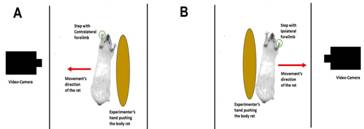
Figure 1 Setup to assess the Steps Adjustment Test. The number of steps with the forelimbs is counted while the rat’s body is gently pushed with the experimenter’s hand on a smooth sheet of stainless steel at a constant speed to a video camera. A) Push to the contralateral side of the lesion. B) Push to the ipsilateral side.
The number of steps performed in each test with each forelimb was counted and averaged to obtain a single-step value for each limb of each rat. Subsequently, these values per rat were averaged to find the group mean per day of evaluation. This procedure was repeated for each experimental phase of SAT (i.e., pre-lesion, post-lesion, and post-implant phases). To evaluate the therapeutic effects of the SiO2 or SiO2-DA reservoir on recovery of symmetry in the use of the forelimbs to perform steps after the lesion, the number of steps performed with the CF was divided between the number of steps with the IF and the result was multiplied by one hundred.
Cylinder Test (CT): This test evaluates the number of contacts that the rat makes with its forelimbs on the wall of a transparent cylinder during postural changes and is analogous to the use of the legs in humans when walking and during postural changes while standing up (17)(18). Since the number of contacts on the cylinder wall represents a more sensitive indicator of the motor deficit due to striatal DAergic depletion and to the response to antiparkinsonian therapy than those made on the floor, only the contacts during vertical exploration were considered in this study. Each rat was placed inside a transparent acrylic cylinder of 30 cm in height and 20 cm in diameter and its activity was videotaped for 10 min. A double mirror was placed behind the cylinder at an appropriate angle to allow registration of the contacts on the cylinder wall, even though the rat had its back to the camera (Figure 2).
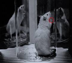
Figure 2 Setup to assess the Cylinder Test. During vertical exploration, the number of contacts on the wall of a clear acrylic cylinder is counted while its activity is videotaped. Two mirrors were placed behind at an angle such the contacts could be recorded whenever the rat had its back to the camera.
The wall contacts were classified as follows: (a) independent contacts on the cylinder wall with the IF or with the CF, and simultaneous or semi-simultaneous contacts with both forelimbs (BF). According to the criteria of Schallert and Tillerson (18): (a) during vertical exploration, the first forelimb used for the initial contact was registered as independent (IF or CF); (b) each lateral movement touching the wall with the forelimb was recorded as independent (IF or CF); (c) when one limb was already making contact with the wall, the posterior placement of the other limb (> 0.4 s) was recorded as simultaneous (BF); (d) if the rat examines the wall laterally, all alternating movements involving the IF and the CF, were recorded as a simultaneous (BF); (e) when one limb maintained contact stationary; while the other limb made small step adjustments, it was recorded as a simultaneous contact (BF); (f) when BF was removed from the wall, the new contact was recorded as independent or simultaneous, as previously described. For a test to be considered valid, the rat needed to make at least twenty contacts on the cylinder wall during the evaluation. When necessary, the rats were encouraged to make contact with the cylinder wall, using the following actions: 1) covering the upper half of the cylinder entrance with a sheet of paper; 2) removing the rat from the cylinder for 30 s and returning it to the cylinder again, 3) turning the room light off and on again several times, and 4) repeatedly making noise on a metal surface or the surface of the cylinder (16)(18)(19). The relative percentage (RP) in the use of IF was calculated as follows: independent use of the IF was divided between the total number of contacts (IF + CF + BF) exhibited by each animal and multiplied by 100. The same relation was employed for RP of CF and RP of BF.
Sequences of the SAT and the CT: The step adjustment test was performed every week on alternate days. To obtain the baseline prelesion (BPRE) values, the test was conducted between days -7 to -3 before the lesion (see Figure 6). To obtain the baseline postlesion (BPOST) values, the test was conducted between days 44 and 48 of the eighth postlesion week. For the groups that received the implants with or without DA, the test was conducted at weeks 12, 16, 20, and 24 PL (corresponding to weeks 4, 8, 12, and 16 PI, respectively) (see Figure 3). To calculate the basal prelesion (BPRE) values, the cylinder test was conducted 1 day before the lesion (day -1) and to obtain the basal postlesion values (BPOST) the test was conducted again on day 50 postlesion (eighth post-lesion week). To assess the therapeutic effect of the DA released from the reservoir of nanostructured silica in the striatum, the test was conducted again at weeks 12 and 24 PL (corresponding to weeks 4 and 16 PI, respectively). (Figure 3)
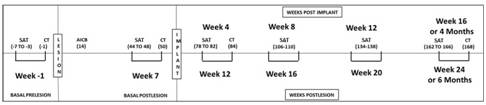
Figure 3 A sequence of the postural adjustment tests performed by the hemiparkinsonian rats. Time of the assays basal pre-lesion, post-lesion, and intrastriatal post-implant of SiO2 or SiO2-DA reservoirs. The numbers in the parentheses are the days of the behavioral tests about the lesion (post-lesion days). Weeks in the upper part are about the moment of placement of intrastriatal implants and those in the lower part are about the day of the lesion of the SNpc. Abbrev.: SAT, Steps Adjustment Test; CT, Cylinder Test; AICB, apomorphine-induced circling behavior.
Histology: At the end of the experiments and 17 weeks after implantation surgery, the test animals were placed under deep anesthesia and their brains were perfused with 200 mL of a cold phosphate buffer solution (PBS, 0.1 M, pH 7.4), followed by 300 mL of cold paraformaldehyde (4%) in PBS, via an infusion system through the ascending aorta. To appraise the extent of DAergic denervation induced with the neurotoxin 6-OHDA, the presence of tyrosine-hydroxylase (T-H), the limiting enzyme in the DA synthesis pathway, was evaluated using immunohistochemistry. The perfused brains were removed, postfixed in 4% paraformaldehyde for 2 h at room temperature, and placed overnight in PBS with 15% sucrose at 4°C. Subsequently, using a microtome (Vibratome, Bannockburn, IL), coronal sections of the brain with a thickness of fifty µm were obtained. The sections included the striatum and SNpc regions. Each section was sequentially placed on multi-well plates containing PBS. Most of the sections were obtained approximately five hundred µm from the rostral pole of the striatum and 6 additional sections more caudal were obtained with a separation of 350 µm, between one and the other. The SNpc was serially cut throughout its total cranio-caudal extension and the sections were stained alternately. The selected sections were incubated for 2 h at room temperature with a blocking solution containing bovine albumin serum (1%), triton-X-100 (0.3%), and sodium azide (0.0125%) in PBS. The sections were incubated for 72 h with polyclonal anti-T-H rabbit antibodies (Chemicon) at a 1:1000 dilution. Then an ABC Vectastain kit (Vector) was used. The sections were washed 3 times with the blocking solution and then incubated for 2 h at room temperature with a mouse biotinylated anti-rabbit IgG secondary antibody, diluted 1:500. After this, the sections were incubated for 2 h with the avidin-peroxidase conjugate and finally revealed in a solution of H2O2 (0.01%) with 3,3'-diaminobenzidine and cobalt chloride as a color enhancer. The immunostained sections were mounted on gelatinized slides, covered with coverslips and subsequently photographed with a digital camera (Olympus DP11) fixed on a microscope (Olympus SZ11).
Statistical analysis: By international regulations for the use of animals in research in Neuroscience, the number of rats used in this study was the minimum required to obtain statistically significant results. To reduce bias, all the observers were blind to the type of rats they were examining when conducting the assessments of the primary outcomes. The results are expressed as Mean ± standard error.
To calculate the average percentage of deterioration after 6-OHDA lesion on the movement of the CF or the IF of the 12 rats that reached the validation criteria of a minimum of 200 contralateral turns after the challenge with apomorphine, the number of steps obtained BPRE was subtracted to the value obtained BPOST and the result was divided by the number of steps in BPRE, and the means obtained of the percentages of IF were compared against CF using an unpaired Student's t-test. To compare the average number of steps of the CF and IF throughout all phases of the study against BPOST, a repeated measures ANOVA test followed by a Dunnett post hoc test was used for each group. To evaluate the efficacy of DA release from the intrastriatal reservoir of nanostructured silica to restore symmetry between the movement of both limbs to BPRE values, the means of the percentage of symmetry obtained at weeks 4, 8, 12, and 16 PI were compared to the values of BPRE, using a repeated measures ANOVA test for each group, followed by a post hoc Dunnett test.
For the cylinder test, to evaluate the effect of SiO2-DA reservoirs implanted in the striatum on the relative percentage of the independent and simultaneous use of the forelimbs to touch the cylinder wall during vertical exploration, the means obtained for each case were compared with a repeated measures ANOVA test followed by a post hoc Dunnett's test. The significance level was set at 0.05. All statistical analyses were carried out using the GraphPad Prism statistical software, version 5.0.
Results
Contralateral circling behavior induced by apomorphine challenge: Two weeks after the lesion, the rats were challenged by apomorphine to evaluate contralateral circling behavior. Twelve rats reached the validation criteria after the challenge with apomorphine with an average of 336.3 ( 43.63 turns/90 min (rank: 200 to 636 turns/90 min).
Steps Adjusting Test (SAT): The twelve rats selected showed a significant reduction in the execution of steps with the CF at BPOST vs . BPRE. All the groups used both the IF and CF to make steps and were symmetrical throughout the BPRE evaluations (days -7 to -3). However, on days 44 to 48 PL, there was a significant deterioration in the CF, but not in the IF. The average percentage of deterioration post-lesion in the number of steps with the CF of the 12 rats evaluated was 81.25 ± 3.09 % (range: de 55.17 a 98.30 %), which was significantly higher ( p < 0.0001) than that observed with the IF.
The animals without implants (N = 4) made an average of 14.65 ± 0.06 steps on BPRE vs . 1.50 ± 0.50 steps on BPOST ( p < 0.0001) (see Figure 4A). Rats with SiO2 reservoir (N = 4) made an average of 14.88 ± 0.06 steps on BPRE vs . 3.81 ± 0.74 steps on BPOST ( p < 0.0001) (see Figure 4B); while the animals with de SiO2-DA (N = 4) reservoir made in average 14.31 ± 0.42 steps at BPRE and it was reduced to 2.25 ± 0.60 steps on BPOST ( p < 0.0001) (see Figure 4C). In rats without an implant and with the SiO2 reservoir, the motor deterioration showed by the CF after the lesion, remained unchanged along with evaluations (until day 166 PL) compared against BPOST values (see Figures 4A and 4B). On the contrary, rats with reservoir SiO2-DA showed a significant increase in the number of steps vs . the BPOST, after the intrastriatal implant, making an average of 2.25 ± 0.60 steps on BPOST vs . 14.00 ± 0.53 steps at day 78 PL ( p < 0.0001), and 14.50 ± 0.10 steps at the day 166 PL ( p < 0.0001), of the week 16 PI (see Figure 4C). The increase in the number of steps with the CF started at day 78 PL and remained stable until the last evaluation of the study, at day 166 PL (week 16 PI). None of the groups showed deterioration in the number of steps with the IF after the lesion, and this behavior remained constant throughout all phases of the study until the last evaluation (see Figures 4A-C).
In BPRE, the symmetry percentage in the use of IF and CF to make steps was almost 100% for all the groups: non-implant, SiO2, and SiO2-DA groups. For the week 12 PL, the groups without implant and with implant without DA had a significant reduction ( p < 0.0001) in the values of symmetry percentage (Lesioned group: average 8.80 ± 2.70%; SiO2 group: average 26.84 ± 8.33%) maintained until the week 24 PL (week 16 PI for the SiO2 group). (see Figures 5A-C)
Only the SiO2-DA group increased its symmetry percentage to almost 100% for week 12 PL (week 4 PI; average 98.85 ± 3.30%) maintained until week 24 PL (week 16 PI). The values of symmetry percentage in the SiO2-DA group didn’t show significant differences against the values obtained in BPRE. The time-beneficial effect on the postural adjustment with the intrastriatal SiO2-DA reservoir in the hemiparkinsonian rats was 16 weeks (4 months), which is equivalent to 10 years in humans (20). (Figure 4)(Figure 5)
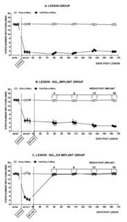
Figure 4 Adjusting steps of the ipsilateral and contralateral forelimbs before and after the lesion, and after the implant. A, Group without implant; B, Group with SiO2 reservoir; C) Group with SiO2-DA reservoir. Abbrev.: BPRE, basal pre-lesion; BPOST, basal post-lesion, before SiO2 or SiO2-DA implants. The asterisk marks the value for both IF and CF (day 48 BPOST) vs. the values of the BPRE (days -7 to -3), and vs. the days PI (days 78 to 168 post-lesion). op < 0.0001; +p < 0.05. Repeated measures ANOVA test, followed by post hoc Dunnett’s test.
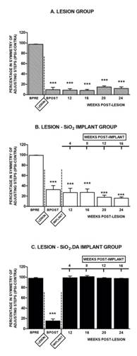
Figure 5 Percentage of symmetry in the execution of steps with contralateral forelimbs. Abbrev. BPRE, basal prelesion; BPOST, basal postlesion. In the upper zone are the weeks after the striatum silica implant without or with DA. In the below zone are the weeks about the nigrostriatal lesion. ***p < 0.0001. Repeated measures ANOVA test followed by post hoc Dunnett’s test.
Cylinder Test (CT): In BPRE, the relative percentages (RP) were 22% for the independent use of each IF or CF and around 58% for the simultaneous or almost simultaneous use of BF. The percentages changed significantly ( p < 0.0001) after the lesion, with an increase in the percentage of the independent use of the IF to around 100%. In comparison, the RP of the independent use of the CF and the simultaneous use of BF were reduced significantly ( p < 0.0001) to around 0%. In the groups of rats without implant or with SiO2 reservoir, these percentages remained unchanged until week 24 PL (week 16 PI). On the other hand, the group with SiO2-DA reservoir showed a significant improvement ( p < 0.0001) in the percentage of the independent use of the CF (average 12.41 ± 4.42%) and the relative percentage in the use of BF (average 27.44 ± 3.05%), on week 16 PI (see Figure 6). (Figure 6)(Figure 7)
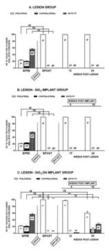
Figure 6 Relative percentage in the use of the forelimbs to make contact with the cylinder wall during vertical exploration. The upper zone shows the weeks post-implant, while in the below zone are the weeks post-lesion. The symbols a, b, and c, above the bars, represent the comparison between the means, during BPOST vs. BPRE of the RPs in the independent (IF or CF) or simultaneous use of BF, respectively. The symbols d, e, and f represent the comparison between the means of the RPs in the independent (IF or CF) or simultaneous use of BF, during BPOST vs. the weeks 12 and 24 PL, respectively. ‡p < 0.0001, +p < 0.001. Repeated measures ANOVA test followed by post hoc Dunnett’s test.
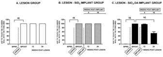
Figure 7 Asymmetry percentage in the use of the forelimbs to touch the cylinder wall during vertical exploration. The symbol a, above the bars, represents the comparison between the means, during BPOST vs. BPRE of the ASPs between the IF and CF. The symbol b, above the bars, represents the comparison between the means of ASPs, during BPOST vs. the weeks 12 and 24 PL, respectively. ‡p < 0.0001. Repeated measures ANOVA test followed by post hoc Dunnett’s test.
None of the hemiparkinsonian rats with SiO2 implants with or without dopamine showed motor oscillations after implants, such as the "On-off" effect or dyskinesias such as those caused by prolonged administration of L-DOPA. (See Figures 7A-C)
Histological analysis: The analysis with immunohistochemistry for T-H showed well-established immune labeling in the left striatum of the SiO2 group (see Figures 8A-B), and SiO2-DA group (see Figures 8D-E), and the left mesencephalon (8C and 8F) for all the lesioned rats. The right striatum and the right side of the mesencephalon showed a lack of immuno-labeling because of the loss of the nigrostriatal pathway with 6-OHDA. In the slices of the right striatum, there was no evidence of DAergic cell regeneration of the nigrostriatal pathway. The presence of reddish-brown particles is remarkable through the SiO2-DA reservoir pathway in the left striatum (see Figures 8D-E) in remarkable contrast to the SiO2 reservoir pathway (see Figures 8A-B) which was translucent. The reddish-brown particles in the SiO2-DA pathway suggest the presence of oxidized molecules, probably DA, which could be oxidized during the brain-fixing process. (Figure 8)
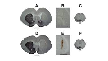
Figure 8 Immunochemistry to T-H in coronal slices of rat’s brain (50 µm) at the level of the striatum and the mesencephalic region. A-B, Coronal slices of rat’s brain at the striatum level with SiO2 reservoir. D-E, Coronal slices of rat’s brain with SiO2-DA reservoir. Scale bar 2 mm. C and F mesencephalic coronal slices with immunochemistry to T-H, where is located the SNpc. B and D are higher magnifications of the areas where are localized the reservoirs. Is noticeable that the pathway of the reservoir with DA has a reddish-brown color, suggesting the presence of oxidizable DA. The slices with immunochemistry to T-H at the striatum and mesencephalic levels (C-E and H-J) clearly show the absence of immune-labeling at the right side of the slices, suggesting the lack of the DAergic cells of the nigrostriatal pathway at that side.
Discussion
The main finding in the present study is that the release of DA from an intrastriatal implanted reservoir in rats with a chronic model of parkinsonism was effective in reversing the deterioration in postural adjustment in a sustained and safe way. The use of SiO2DA did not cause adverse effects or dyskinesias as happens with the systemic administration of drugs such as L-DOPA.
The absence of a beneficial effect on motor activity in parkinsonian rats without implant or with the SiO2 reservoir reinforces the idea that it is the DA released from the intrastriatal reservoir (see Figure 9) that produces the beneficial effect on the postural adjustment of hemiparkinsonian animals and not the sole presence of the silica matrix, the mechanical alterations caused during the stereotactic placement of the implant or the periodic repetition of the adjusting step test.
Injection into the right side of the midbrain of the 6-OHDA neurotoxin resulted in the massive death of the DAergic neurons of the SNpc. This produced a significant reduction in the steps performed with the contralateral forelimb during the adjusting step test, without the involvement of the ipsilateral limb, resulting in a marked asymmetry between both forelimbs, according to previous studies (16). The asymmetry in the use of the forelimbs to execute steps observed after the lesion of the SNpc of the right side of the midbrain remained unchanged throughout the study period during the step adjustment evaluations in the rats without implant and until the week 16 PI in rats with the implant of SiO2 without DA. Only in rats with the intrastriatal SiO2-DA implant, the deterioration in the number of steps with the CF was significantly reduced from the first postimplant evaluation (week 4 PI), with recovery in symmetry in the number of steps between the IF and CF, around 100%, and without significant difference with the values of the BPRE. Once symmetry was reached in the execution of steps with the IF and CF, it remained stable, that is without oscillations, during the following evaluations until the week 16 PI, suggesting the controlled release of DA within the striatum (see Figure 9) during all this time (4 months), which is equivalent to 10 years in humans (20).
The histological analysis confirmed the correct placement of the SiO2-DA reservoir which was found in the dorsal region of the striatum. The entry pathway of the SiO2-DA reservoir in the striatum showed reddish brown particles which suggested the presence of oxidizable DA in this area, while on the contrary, the entry pathway of the SiO2 reservoirs was translucent. Since during the perfusion process, oxidating substances were used to fix the tissue and during the brain cutting process and the assembly of the cuts obtained there was exposure to the environment, the DA molecules that were still conserved within the reservoir could have been oxidized. This finding supports that the DA molecules were able to remain functional within the reservoir before the process of euthanizing the rats and perfusing the brains. This agrees with in vitro studies that evaluated the stability of DA within reservoirs of nanostructured silica (4). Furthermore, in the post-mortem analysis of the brains of the hemiparkinsonian rats with the use of immunohistochemistry for tyrosine-hydroxylase (T-H), a lack of immunolabeling to the T-H enzyme is observed in the striatum and the SNpc on the right side of the brain. This means that in the restoration of motor symmetry in the adjusting step test, there was no participation of cell regeneration, as demonstrated by the lack of immunolabeling of T-H in the right side of the striatum and the SNpc, confirming the absence of this enzyme, and therefore, of viable DAergic neurons.
The results of this study showed that the unilateral lesion of the nigrostriatal DAergic pathway induced a significant reduction in the independent use and in the simultaneous use or association with the IF to contact the wall of a transparent cylinder during vertical exploration, considering analogous the use of human legs during postural changes, by previous studies (17)(18). In animals without implants or with SiO2 reservoirs, the deterioration in the relative percentage of the independent use of the CF or in association with the use of the IF to contact the cylinder wall remained unchanged until week 24 PL (week 16 PI, respectively). On the contrary, in animals with SiO2-DA reservoirs, the relative percentage in the independent use of the CF or in association with the IF (both limbs) to contact the cylinder wall showed a significant increase in the week 16 PI evaluation, although no significant effect was observed yet in the week 4 PI.
The difference in sensitivity between the postural adjustment tests to the treatments was also observed in a previous study with systemic administration of caffeine (17). In that study was observed that the adjusting step test is more sensitive to the beneficial effect of the treatment than the cylinder test. For example, with the daily administration of caffeine at a dose of 3 mg/kg for 3 weeks, the maximum effect on the execution of steps of the CF was observed from the first week of treatment, while in the cylinder test, there was only a significant increase in BF up to the second week of treatment with no change in the independent use of the CF. Unlike the study with systemic administration of caffeine at the dose of 3 mg/kg, it’s important to note that in the present study, the rats with a SiO2-DA reservoir implant didn’t present a significant change in the RP of independent use of CF or the RP of BF to touch the cylinder wall at week 4 PI. Until week 16 PI, the RP in the independent use of the CF had a significant increase against the BPOST values, although significantly lower than that obtained in BPRE.
Together, our results showed a prolonged benefit with the use of an intrastriatal implant of SiO2-DA reservoir in hemiparkinsonian rats, with a sustained reduction of postural instability. The hypothesis that the prolonged improvement in postural adjustment in the hemiparkinsonian rats was due to the prolonged and stable release of DA intrastriatal, is supported by the lack of motor improvement in the animals with the intrastriatal implant of SiO2 reservoir without DA, while the motor improvement in the postural adjustment tests in rats with the SiO2-DA reservoir maintained until the fourth month of evaluation, and there was the presence of oxidizable particles present in the perfused brains of these rats even at 17 weeks PI. (Figure 9)
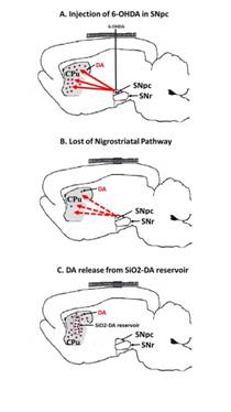
Figure 9 Dopamine release in the striatum of hemiparkinsonian rats since SiO2-DA reservoirs. A. Lesion of SNpc with 6-OHDA, B. Loss of DAergic nigrostriatal pathway, C. Release of DA in the striatum since SiO2-DA reservoir. Abbrev. SNpc, Substantia nigra pars compacta; SNr, Substantia nigra reticulata; CPu, Caudado-Putamen nucleus (Striatum), 6-OHDA, 6-hidroxydopamine; DA, dopamine.
Accordingly, the stability of the response in the adjusting step test at weeks 4, 8, 12, and 16 PI showed an intrastriatal controlled release of DA. In the present study, the animals didn’t show behavioral alterations outside the predicted by the hemiparkinsonism model. Neither adverse effects due to prolonged intracerebral DA release were observed, such as the “on-off” phenomenon nor motor abnormalities such as dyskinesias. Side effects are frequently induced by prolonged systemic administration of L-DOPA in patients with PD (2) and in hemiparkinsonian rats (21)(22). The present study suggests that the use of SiO2-DA reservoirs implanted in the striatum may also be effective in the reduction of akinesia and in increasing postural stability in patients with PD. Although more studies are required to ensure this proposal, the results obtained in this study are promising.
Conclusion
This study demonstrated that the nanostructured silica reservoirs with DA implanted in the striatum of hemiparkinsonian rats produced a significant improvement in postural adjustment with prolonged recovery in the use of the CF to execute steps, as well as in the RP in the independent use of the CF, and BF for contacting the cylinder wall. We didn’t observe rejection to the reservoir or development of dyskinesias or any other adverse effect during the total time of the study. The results of this study demonstrate that intrastriatal implants with DA have the potential to be used as a safe and effective way of sustained release of drugs in patients with PD. However, further studies are necessary not only to assess the prolonged effects of SiO2-dopamine implants but also to evaluate possible toxicity effects of the SiO2 implant itself beyond the scope of this study

