SciELO - Scientific Electronic Library Online

 
vol.33 número1Tratamiento De Migraña En EmbarazoParaparesia Flácida Subaguda Progresiva Y Meningitis Carcinomatosa Como Forma De Presentación De Recaída Tardía De Leucemia Linfoblástica Aguda De Estirpe Linfoide B BCR-ABL/Phi+. índice de autoresíndice de materiabúsqueda de artículos
Home Pagelista alfabética de revistas  

Servicios Personalizados

Revista

Articulo

Indicadores

Links relacionados

  • No hay articulos similaresSimilares en SciELO

Compartir


Revista Ecuatoriana de Neurología

versión On-line ISSN 2631-2581versión impresa ISSN 1019-8113

Rev Ecuat Neurol vol.33 no.1 Guayaquil abr./jun. 2024

https://doi.org/10.46997/revecuatneurol33100097 

Review Article

Neuropsychological And Brain Atrophy Pattern Characterization In Patients With Stroke Related To COVID Infection: A Systematic Review.

Caracterización Neuropsicológica Y Del Patrón De Atrofia Cerebral En Pacientes Con Ictus Relacionado A COVID: Una Revisión Sistemática.

Ivo Bousek-Barraza1 

Constanza Rodríguez-Jorquera1 

Melissa Carrasco-Hermosilla1 

Teresa Julio-Ramos2 

José Jorquera-Araya3 

Igor Cigarroa4 

David Toloza-Ramirez5 

1Universidad Andres Bello. Santiago, Chile.

2Universidad de Valparaiso. Valparaiso, Chile.

3Universidad Pedro de Valdivia. Santiago, Chile.

4Universidad Católica Silva Henríquez, Facultad de Salud, Escuela de Kinesiología. Santiago, Chile.

5Universidad Andres Bello, Faculty of Rehabilitation Sciences, School of Speech Therapy, Santiago, Chile. <david.toloza@me.com>


Abstract

Introduction:

The high mortality rates in patients with COVID infection have been the main focus of interest in clinical and research practice. COVID infection has also been characterized by its respiratory consequences; however, several studies have reported neurological manifestations in this clinical population (e.g., stroke). From a neurological perspective, patients with stroke related to COVID infection remain challenging as well. No systematic reviews have yet focused on neuropsychological and neuroanatomical correlate profiles.

Aim:

To characterize the neuropsychological profile and the pattern of brain areas affected in adult and older adults with stroke related to COVID infection.

Methods:

The present study followed the PRISMA guidelines and included studies from the PubMed, Scopus, and Web of Sciences databases, published between January 2020 and September 2022.

Results:

7 articles were included in this systematic review for critical analysis. Ischemic stroke was the most reported in patients. Results suggest that the most reported neuropsychological declines are orientation, attention, memory, executive function, visuoconstructive skills, and language. The pattern of brain areas also considers right lesions in cortical and subcortical regions.

Conclusion:

In patients with COVID-related strokes, lesions in the left hemisphere or bilateral are common, together with the expression of aphasia. Likewise, our findings reflect only ischemic patients' cognitive and brain features. Therefore, results must be interpreted carefully.

Keywords: Stroke; COVID-19; Diagnosis; Neuropsychological Assessment; Neuroanatomical Correlate; Atrophy pattern.

Resumen

Introducción:

Las altas tasas de mortalidad por COVID, han sido el foco de interés en la práctica clínica e investigativa. La infección por COVID ha sido caracterizada principalmente por sus repercusiones respiratorias; sin embargo, se han reportado manifestaciones neurológicas en esta población (p.e., ictus). Desde una perspectiva neuropsicológica, el ictus relacionado a COVID sigue siendo un desafío en la actualidad. De hecho, existe carencia de estudios enfocados en el perfil neuropsicológico y el correlato neuroanatómico en estos pacientes.

Objetivo:

Caracterizar el perfil neuropsicológico y el patrón de áreas cerebrales afectadas en adultos y personas mayores con ictus relacionado a COVID.

Metodología:

La presente revisión siguió los lineamientos PRISMA, utilizando las bases de datos PubMed, Scopus, y Web of Sciences, incluyendo estudios publicados entre Enero 2020 y Septiembre 2022.

Resultados:

7 artículos fueron incluidos para análisis, siendo el ictus isquémico el más reportado. Los resultados sugieren declives neuropsicológicos en orientación, atención, memoria, función ejecutiva, habilidades visuoconstructivas, y lenguaje. Asimismo, el patrón de áreas cerebrales afectadas también considera lesiones derechas, corticales y subcorticales.

Conclusión:

En pacientes con ictus relacionado a COVID, son comunes las lesiones cerebrales izquierdas o bilaterales, junto a la manifestación de afasia. Nuestros hallazgos sólo reflejan características cognitivas y cerebrales de pacientes con ictus isquémico, por lo cual estos resultados deben ser interpretados con cautela.

Palabras claves: Ictus; COVID-19; Diagnóstico; Evaluación neuropsicológica; Correlato neuroanatómico; Atrofia cerebral.

Introduction

COVID infection is characterized by severe acute respiratory syndrome. During the first year after its debut, it became the leading cause of hospitalization (1). This disease has infected more than 4 billion people worldwide after two years and is responsible for more than 5 million deaths (2)(3). Carod-Artal (4) suggests that the mortality rate associated with COVID infection is around 8%, with adults and older adults with chronic diseases most affected (e.g., immunosuppression).

The most reported symptoms of COVID infection are pneumonia, odynophagia, cough, and nasal congestion. After the onset, they rapidly progressed to complex respiratory symptoms with a high risk of mortality (4)(5). However, several studies highlight the neuroinvasive capacity of the virus and the development of neurological symptoms (1)(4)(5)(6).

Coagulopathy associated with COVID infection leads to systemic thromboembolism. Venous and arterial thromboembolism increases the risk of a neurological event (e.g., stroke) (7). Yamakawa et al. (7) emphasized that people with COVID infection have shown a greater predisposition to developing ischemic strokes. Thieme et al. (8) highlighted that the importance of stroke and its consequences in the population due to COVID infection is characterized by cognitive and motor effects and also by long-term disabilities.

Several studies have reported the neuropsychological repercussions of strokes, emphasizing risk factors (9)(10)(11)(12)(13). Stroke related to COVID infection cases has yet to be profiled from a neuropsychological and neuroanatomical perspective, though. Although recent investigations (14)(15)(16)(17)(18)(19) have reported cognitive associations, these are limited to highlighting cognitive impairment but do not mention the cognitive domains affected and the severity. These studies’ results also suggest that neuropsychiatric symptoms, physical repercussions, and fatigue are frequent. Studies based on neuroimaging techniques have reported dysfunctions in the cingulate cortex and brainstem (16)(20); however, few studies established associations with memory, attention, executive function, or language decline (21)(22)(23)(24)(25).

Despite the significant impact of COVID infection and its increasing interest in clinical and research practice, there have not yet been any reviews that characterize stroke cases related with this infection from a neuropsychological and neuroanatomical perspective. It is thus essential to synthesize the existing evidence of the neurological repercussions of COVID infection. Identifying neuropsychological and neuroanatomical deficits would contribute to pertinent neurorehabilitation programs, which decrease functional disability (26)(27)(28).

Based on the above, our systematic qualitative review aims to characterize the neuropsychological profile and atrophy pattern of the brain areas affected by strokes related with COVID infection in adults and the elderly. Even though cases of COVID infection have fallen worldwide, the information provided will be vital to improving the assessment and intervention process in clinical practice.

Methodology

This systematic qualitative review followed PRISMA guidelines (29) and was previously registered on the PROSPERO repository (PROSPERO 2022: CRD42022363907).

Sources and search strategy

The search for bibliographic references considered the PubMed, Scopus, and Web of Science databases, including articles published between January 2020 and September 2022. The search syntax was as follows: [adult OR adults] AND [aging OR elderly] AND [COVID-19 OR SARS-CoV-2 infection OR coronavirus infection] AND [stroke OR cerebral stroke OR ischemic stroke OR cerebral hemorrhage] AND [cognition OR cognitive decline OR cognitive impairment] AND [neuropsychological assessment OR neuroimaging OR brain imaging]. All search terms were adapted to each database (Appendix 1).

Study selection and eligibility criteria

The following inclusion criteria were applied: a) adults and older adults from 30 to 75 years, b) no clinical history of previous stroke, c) analytical observational studies of diagnostic tests published in English between January 2020 and September 2022, and d) studies reporting neuropsychological and neuroimaging assessment. Exclusion criteria were: a) editorial articles, systematic reviews, protocols or theses, and b) patients with a dementia or mild cognitive impairment diagnosis.

Data extraction

To eliminate duplicate articles, we imported the studies to the Rayyan software (30). Subsequently, three reviewers (I.B-B., M.C-H., and C.R-J) applied the inclusion and exclusion criteria independently. In case of doubt, the research team retrieved articles’ full texts when decisions could not be made based on the title and abstract. Based on his experience (D.T-R), a fourth investigator participated in the consensus selection of those studies presenting discrepancies (15%) regarding their inclusion.

Risk of bias and quality assessment tool

To assess the bias risk and the articles' methodological quality, we used the QUADAS-2 tool (31). For the bias analysis, four key domains were assessed: patient selection, index test, reference standard, and flow and timing domain. Methodological quality was evaluated based on patient selection, index test, and reference standard domain. Finally, high, low, and unclear criteria classified each included study.

Data synthesis strategy

The general characteristics of the studies, such as year of publication, population, mean age, type of stroke, time from neurological symptoms, COVID severity/phase, and Intensive Care Unit admission, are in Table 1. Table 2 presents the principal neuropsychological declines and neuroanatomical findings in patients with stroke related to COVID infection, and the main instruments/measures used. It is important to mention that the changes in each neuropsychological capacity, shown in Table 2, were classified into increase or decrease based on the score reported for the MoCA test.

Results

Literature search

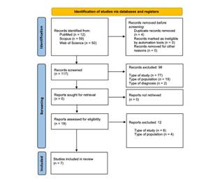
The article selection process is illustrated in the PRISMA flowchart (see Fig. 1) (29). We identified titles and abstracts of 121 articles, considering four duplicates. After applying the inclusion/exclusion criteria to the 117 studies screened, 98 articles were excluded. 19 full texts were analyzed to assess their eligibility; however, after the analysis, eight articles were excluded. Finally, seven articles were included for review. (Figure 1)

Figure 1 Flow Diagram PRISMA search and selection articles. 

General characteristics of the articles

Table 1 summarizes the general characteristics of the seven studies (32)(34)(35)(36)(37)(38) included. For publication years, 2021 stands out with three articles, followed by 2020 and 2022 with two studies, respectively. The total sample was 1,081 adult and older adult patients, with 664 cases of patients with COVID infection-related strokes (mainly ischemic stroke). For age and gender variables, the mean age for stroke patients related to COVID infection was 60.6 years. Most patients were male (60%), for both stroke-related or not with COVID infection. The timeframe for expressing neurological symptoms related to COVID infection was 2 to 12 days. The most reported COVID infection severity was mild-moderate, and the acute phase was reported in 6 articles. Finally, regarding the need for Intensive Care Unit admission, results suggest that 57% of the articles reported no need. (Table 1)

Table 1 General study characteristics. 

First Author Participants Sex Sex Mean Age sCOVID-inf (years, SD) Age range Type of stroke (%) Time of neurological symptoms (days) COVID infection severity (%) a COVID infection phase ICU admission
sCOVID-inf/ tCOVID-inf ♀ ♂ ♀ ♂ (years)
% tCOVID-inf % sCOVID-inf 30-49 50-69 70 + Is Hem Throm Mil-Mod Sev-Cri Yes No
Meppiel et al. (2021) (32) 57/222 38.7 61.3 40.4 59.6 65 (-) - 92 - 8 12 65 35 Acute phase -
Chaumont et al. (2022) (33) 18/60 40 60 - - 66 (-) - 94 6 - 5 57 43 Long COVID infection -
Chougar et al. (2020) (34) 66/73 34.2 65.8 - - 58.5 (15.6) 65 31 4 6 52 48 Acute phase -
Shahjouei et al. (2021) (35) 432 stroke patients associated with COVID-19 42.4 57.6 42.4 57.6 65.7 (15.7) 75 21 4 3-5 - - Acute phase -
van Lith et al. (2022) (36) 70/200 - - - - - 100 - - 2 - - Acute phase -
Sabayan et al. (2021) (37) 15 stroke patients associated with COVID-19 20 80 20 80 65 (15.6) 93 7 - 7 27 73 Acute phase -
Ashrafi et al. (2020) (38) 6 stroke patients associated with COVID-19 50 50 50 50 43.5 (7.42) - 100 - - 2-6 83 17 Acute phase -

*Notes. ✓: Specified. -: Not specified. sCOVID-inf: stroke patients related to COVID infection. tCOVID-19: total of patients related to COVID infection included in the study. SD: Standard deviation. Is: Ischemic. Hem: Hemorrhagic. Throm: Thrombotic. Mil: Mild. Mod: Moderate: Sev: Severe. Cri: Critical. ICU: Intensive Care Unit. a: According to National Institute of Health guidelines.

Neuropsychological and Neuroanatomical perspective

Table 2 reports that neuropsychological assessment was based mainly on the MoCA test and the criteria established in NIHSS. The declines reported in patients with COVID infection-related strokes include alterations in spatial and temporal orientation, attention, memory, visuoconstructive skills, and linguistic skills (such as expressive, comprehensive, naming, and semantic and phonological fluency).

The most reported neuroimaging techniques were CT and MRI. Cortical involvement comprises the frontal, temporal, parietal, and occipital lobes, although involvement of the corpus callosum, brainstem, and subcortical areas is also reported. The damage pattern is limited to not only the left but also to bilateral affection. (Table 2)

Table 2 Neuropsychological and Neuroanatomical characterization. 

Ref. Neuropsychological characterization Neuroanatomical characterization
Instruments/Measures Neuropsychological findings Instruments/Measures Hemisphere localization Neuroanatomical findings (stroke localization)
(32) GNE (: MM, T-S-O, VCA, PF, RE, SS, Syn-Gram abilities (: AN MRI, CT, CSF, EEG, ENM L, BI Frontal, parietal, occipitotemporal and cerebellar areas.
(33) mRS, EQ-5D-3L, CDS, MoCA, HADS (: MM, DR, EF, AT, OR, VSA, AC, RE, PF CT NE Subcortical areas.
(34) Clinical data collected by neurologists. (: AT, T-S-O MRI, EEG, CSF, CT NE Corpus callosum, basal ganglia, sustantia nigra, globus pallidus, and the nigrostriatal pathway.
(35) NIHSS (: T-S-O, AT, CS, ES, PF, SF, NAM MRI, CT, TOAST NE Basal ganglia, superior sagittal sinus, and sigmoid sinuses.
(36) HADS, PCFS, mRS, MoCA, TICS (: MM, VCA, AT, EF, SF, PF MRI EC L, BI The results suggest that the damage is mainly in cortical regions; however, the affected brain areas are not deepened.
(37) NIHSS, mRS (: T-S-O, LA, MM, AT CT MRI L, BI Striatum, brainstem, temporal, parietal, and occipital lobes.
(38) NIHSS (: LA, CS, ES, Syn-Gram abilities, SF, PF, NAM, AT, MM, EF. (: AN ECG CT TCCS EC L, BI Basal ganglia, caudate, lentiform nucleus, and anterior horn of the internal capsule.

*Notes. Ref: References. : Decrease. : Increase. GNE: General neuropsychological evaluation. mRS: Modified Rankin Scale. EQ-5D-3L: 3-level version of the EuroQol five-dimension scale. CDS: 35-item version of Cognitive Difficulties Scale. MoCA: Montreal Cognitive Assessment. HADS: Hospital Anxiety and Depression Scale. NIHSS: National Institutes of Health Stroke Scale. PCSF: post-COVID functional scale. TICS: Telephone Interview for Cognitive Status. MM: Memory. T-S-O: Temporal and spatial orientation. VCA: Visuoconstructive abilities. PF: Phonological fluency. RE: Repetition. SS: Spontaneous speech. Syn-Gram: Syntactic-Grammatical. AN: Anomia. DR: Delayed recall. EF: Executive function. AT: Attention. OR: Orientation. VSA: Visuospatial abilities. AC: abstraction capacity. CS: Comprehensive skills. ES: Expressive skills. SF: Semantic fluency. NAM: Naming. LA: Language abilities. MRI: Magnetic resonance imaging. CT: Computed tomographic. CSF: Cerebrospinal fluid. EEG: Electroencephalogram. ENM: Electroneuromyography. TOAST: Trial of ORG 10172 in Acute Stroke Treatment. EC: Echocardiography. ECG: Electrocardiogram. TCCS: transcranial color-coded duplex ultrasonography. NE: Not specified. L: Left. BI: Bilateral.

Risk of bias and methodological quality of the articles

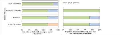
The analysis of the seven articles with QUADAS-2 (31) appears in Fig. 2. Regarding the risk of bias analysis, 72% were classified as low risk in patient selection, while 28% had uncertain risk in the test index and reference test domains. 86% were considered low risk in flow and timing. On the other hand, in the methodological quality assessment, 72% of the studies were classified as low risk for patient selection and test index. 100% were classified as low risk in the reference test domain. (Figure 2)

Figure 2 Risk of bias and methodological quality. 

Discussion

The present review aimed to characterize adults and older adult patients with stroke related to COVID infection, using neuropsychological and neuroanatomical perspectives. Our findings suggest that orientation, attention, memory, visuoconstructive abilities, and language experienced a reduction in their capacity. On the other hand, the left-brain and bilateral areas most affected are frontal, temporal, parietal, and occipital, in addition to the corpus callosum and subcortical regions.

Stroke and prevalence

The prevalence of stroke and its association with gender variables has been getting a grownup interest in research practice. Studies report opposites outcomes since they suggest no gender differences (39), while other founding suggests higher prevalence in women (40)(41). In line with our findings, Mao et al. (42) indicate that stroke is more common in men. Although the evidence shows disagreement, there is a consensus that the prevalence in men and women depends on age. Studies propose that stroke is frequent between 60-64 years, which is consistent with our results in patients with COVID infection-related strokes who report a mean age of 60.6 years (43)(44).

Several studies have reported a considerable increase in strokes among people under 55 years in recent decades (45)(46)(47). Putaala et al. (48) performed a multicenter study in Europe, finding that the incidence of stroke in women surpasses the incidence of men for age groups under 35 years, while in the age group between 35 and 50, the highest incidence in men prevailed. On the other hand, the evidence highlights that female hormones could play a protective role in the expression of neurological events (even in preclinical models), which could justify the small number of women in our analyzed sample (49)(50)(51)(52)(53).

Ischemic and hemorrhagic stroke

The type of COVID infection-related stroke appearing in our results concords with the current evidence, which reports a high prevalence of ischemic stroke (54)(55)(56)(57). An observational study (44) suggests that 31% of COVID infection patients manifested thrombotic complications, followed by deep vein thrombosis and ischemic stroke. It should also be noted that our results show an important number of hemorrhagic strokes in older adult. The subjects’ age range has been associated with a higher risk of developing mild cognitive impairment or dementia (58)(59)(60)(61). On the other hand, a previous meta-analysis (62) suggests that in stroke patients related to COVID infection, the damage shows greater extension and severity, being the most frequent affliction in large vessels and multiple territories.

Neuroanatomical correlates in stroke

Previous studies highlight that neuroimaging techniques such as MRI and CT are essential to diagnostic accuracy and differentiation between the different types of strokes in the acute stage (63)(64)(65)(66). Our findings report that in patients with COVID infection-related strokes, the damage can affect both cortical and subcortical areas. Indeed, studies have reported that subcortical regions such as the putamen, globus pallidus, and insula also tend to experience significant damage (67)(68).

Case studies in patients with COVID infection-related strokes agree with our brain areas reported. Avula et al. (54) studied four patients with COVID infection and stroke, reporting a significant decrease of white and gray matter in the parietal and occipital lobes, along with predominantly left frontotemporal and corticospinal tract hypodensity. Their results highlight that cognitive decline tends to affect these patients’ language; however, they establish an association between this neuropsychological decline and motor repercussions with damage to the basal ganglia and internal capsule. These data mesh with the findings of another COVID infection case study (69), but both left and right hemisphere damage was reported in the five patients analyzed.

Stroke and its neuropsychological implications

It is postulated that typical post-stroke cognitive domain deficits depend on the injury type, extension, and localization (70). Executive function, working memory, and information processing speed are the most frequent cognitive domains affected, with more severe deficits in older adults (71)(72)(73). Our results in patients with stroke related to COVID infection, similar to other studies, highlight the compromise in orientation, attention, and language (74)(75)(76)(77).

Sharifi-Razavi et al. (78) presented 3 cases of stroke secondary to COVID infection, suggesting that declines in orientation, memory, and language (e.g., aphasia) are highly prevalent in these patients. This is consistent with our findings and the reports from post-COVID patients without stroke. However, in their follow-up study, Mattioli et al. (79) suggest no significant neuropsychological impairments in patients with stroke related to COVID infection.

Aphasia classically tends to be affected in typical post-stroke patients, with mainly left hemisphere damage (80)(81). However, our study also suggests bilateral affection in patients with stroke related to COVID infection, which differs from reports among the non-COVID-infected population. This could indicate possible right hemisphere impacts in this group of patients, perhaps in cases of long COVID. This makes it necessary to investigate possible linguistic disorders secondary to atypical damage in the non-dominant cerebral hemisphere, such as the right hemisphere, which tends to be not associated with linguistic deficits (82).

Neuropsychological assessment

We reported that the NIHSS scale and MoCA test were mainly used to register deficits in patients with stroke related to COVID infection. NIHSS has been widely suggested in the literature, highlighted as an attentive scale for evaluating post-stroke patients related to COVID infection (40)(54)(69)(83)(84)(85). Although there is consensus on the use of this scale, it does not allow an exhaustive evaluation of the different cognitive-linguistic domains in post-stroke patients.

Our results report a wide use of the MoCA test as well. Chiti and Pantoni (86) highlight that it is not frequently used in evaluating stroke patients, but is common in people with mild cognitive impairment or dementia. Despite this, it has excellent psychometric properties to detect cognitive alterations. It has shown good sensitivity and specificity indexes to identify cognitive declines in post-stroke patients (87)(88)(89). Khaw et al. (90) suggest that the MoCA test should be used cautiously in post-stroke people, because it does not consider either a detailed neuropsychological assessment or an exhaustive language evaluation. So far, few studies (91)(92) have focused on language impairment in patients with COVID infection. Those studies highlight that lexical fluency and naming performance could affect these patients; however, they suggest that sociocultural language differences must be considered during assessment.

Limitations

Our systematic review does not present a meta-analysis, focusing only on qualitative analysis. Likewise, we only considered three databases for articles search; therefore, our findings must be interpreted carefully. Moreover, most of the studies defined cognitive impairment using only the MoCA test and NIHSS; however, for some papers, symptoms were reported by clinicians only. Finally, the neuropsychological assessment in all the studies included does not report test scores. Neuropsychological deficits in patients with stroke related to COVID infection were thus performed from a general perspective (qualitative interpretation of the main findings in each article).

Clinical contributions

A previous review by Vanderlind et al.(93) focused on patients' neuropsychological and neuropsychiatric sequelae related to COVID infection; however, they did not establish a neuroanatomical correlation with neuropsychological symptoms. Wilson et al. (94) highlight the importance of neuropsychological performance on stroke patients related to COVID infection, suggesting the importance of accurate detection for appropriate treatment. To our knowledge, this is the first systematic review concerning COVID infection-related strokes to characterize this clinical population from a neuropsychological and a neuroanatomical perspective. Our results provide relevant information for clinical practice, suggesting a generalized profile for implementing specific intervention programs in the acute stage of patients with COVID infection-related strokes and post-COVID patients without stroke. These interventions will reduce functional disability index value and will contribute to delaying the progression of vascular dementia.

Future directions

Although our study focused on the neuroanatomical correlation from a structural damage perspective, future research should consider functional MRI to determine the pattern of brain connectivity affected in patients with COVID infection-related strokes and its implications in the chronic stage and long COVID cases. Finally, the assessment process must establish unified cognitive-linguistic protocols for these patients and the general stroke population.

Conclusions

Results show similar cognitive deficits to those reported in typical strokes. Memory, orientation, attention, and executive function decline are the most common symptoms of COVID infection-related stroke patients, which concords with reports from the general stroke population. Aphasia is also reported, but not only limited to left-side lesions. Cortical and subcortical involvement is postulated in these patients as well, and alterations are mainly presented in the left hemisphere or bilaterally in the clinical population with ischemic stroke.

Finally, it must be noted that the neurorehabilitation process should be performed in the acute stage. Early intervention in this population is critical, considering their susceptibility to developing dementia secondary to stroke.

References

Requena M, Olivé-Gadea M, Muchada M, García-Tornel Á, Deck M, Juega J, et al. COVID-19 and Stroke: Incidence and Etiological Description in a High-Volume Center. J Stroke Cerebrovasc Dis 2020; 29(11):105225. https://doi.org/10.1016/j.jstrokecerebrovasdis.2020.105225 [ Links ]

Kwok HF. The significance of advanced COVID-19 diagnostic testing in pandemic control measures. Int J Biol Sci 2022; 18(12):4610-4617. http://doi.org/10.7150/ijbs.72837 [ Links ]

Ritchie K, Chan D, Watermeyer T. The cognitive consequences of the COVID-19 epidemic: collateral damage? Brain Commun 2020; 2(2):fcaa069. https://doi.org/10.1093/braincomms/fcaa069 [ Links ]

Carod-Artal FJ. Complicaciones neurológicas por coronavirus y COVID-19. Rev Neurol 2020 ; 70(9):311-322. https://doi.org/10.33588/rn.7009.2020179 [ Links ]

Desforges M, Coupanec AL, Dubeau P, Bourgouin A, Lajoie L, Dubé M, et al. Human Coronaviruses and Other Respiratory Viruses: Underestimated Opportunistic Pathogens of the Central Nervous System? Viruses 2019; 12(1):14. https://doi.org/10.3390/v12010014 [ Links ]

Zapa-Pérez N, Moreno-Vargas E, Sierra-Villalobos V, Castro-Salcedo C, Mesa-Santamaria K, Jímenez-Monsalve CA. Neurological symptoms associated with SARS-CoV-2 infection in a tertiary hospital in Bogota, Colombia. Neurol Perspect 2022; 2(2):67-72. https://doi.org/10.1016/j.neurop.2021.12.003 [ Links ]

Yamakawa M, Kuno T, Mikami T, Takagi H, Gronseth G. Clinical Characteristics of Stroke with COVID-19: A Systematic Review and Meta-Analysis. J Stroke Cerebrovasc Dis 2020; 29(12):105288. https://doi.org/10.1016/j.jstrokecerebrovasdis.2020.105288 [ Links ]

Thieme H, Morkisch N, Mehrholz J, Pohl M, Behrens J, Borgetto B, et al. Mirror therapy for improving motor function after stroke. Cochrane Database Syst Rev 2018; 7:CD008449. https://doi.org/10.1002/14651858.CD008449.pub3 [ Links ]

Einstad MS, Saltvedt I, Lydersen S, Ursin MH, Munthe-Kaas R, Ihle-Hansen H, et al. Associations between post-stroke motor and cognitive function: a cross-sectional study. Bmc Geriatr 2021; 21:103. https://doi.org/10.1186/s12877-021-02055-7 [ Links ]

Knight-Greenfield A, Nario JJQ, Gupta A. Causes of Acute Stroke A Patterned Approach. Radiol Clin N Am 2019; 57(6):1093-1108. https://doi.org/10.1016/j.rcl.2019.07.007 [ Links ]

Levine DA, Galecki AT, Okullo D, Briceño EM, Kabeto MU, Morgenstern LB, et al. Association of Blood Pressure and Cognition after Stroke. J Stroke Cerebrovasc Dis 2020; 29(7):104754. https://doi.org/10.1016/j.jstrokecerebrovasdis.2020.104754 [ Links ]

Murray LL. Attention and Other Cognitive Deficits in Aphasia: Presence and Relation to Language and Communication Measures. Am J Speech-lang Pat 2012; 21(2):S51-S64. https://doi.org/10.1044/1058-0360(2012/11-0067) [ Links ]

Rostamian S, Mahinrad S, Stijnen T, Sabayan B, de Craen AJM. Cognitive Impairment and Risk of Stroke. Stroke 2018; 45(5):1342-1348. https://doi.org/10.1161/STROKEAHA.114.004658 [ Links ]

Ceban F, Ling S, Lui LMW, Lee Y, Gill H, Teopiz KM, et al. Fatigue and cognitive impairment in Post-COVID-19 Syndrome: A systematic review and meta-analysis. Brain Behav Immun 2022; 101:93-135. https://doi.org/10.1016/j.bbi.2021.12.020 [ Links ]

Evans RA, McAuley H, Harrison EM, Shikotra A, Singapuri A, Sereno M, et al. Physical, cognitive, and mental health impacts of COVID-19 after hospitalisation (PHOSP-COVID): a UK multicentre, prospective cohort study. Lancet Respir Med 2021; 9(11):1275-1287. https://doi.org/10.1016/S2213-2600(21)00383-0 [ Links ]

Hugon J, Msika EF, Queneau M, Farid K, Paquet C. Long COVID: cognitive complaints (brain fog) and dysfunction of the cingulate cortex. J Neurol 2022; 269:44-46. https://doi.org/10.1007/s00415-021-10655-x [ Links ]

Stefano GB. Historical Insight into Infections and Disorders Associated with Neurological and Psychiatric Sequelae Similar to Long COVID. Med Sci Monit 2021; 27:e931447-1-e931447-4. http://doi.org/10.12659/MSM.931447 [ Links ]

Xu Y, Zhuang Y, Kang L. A Review of Neurological Involvement in Patients with SARS-CoV-2 Infection. Med Sci Monit 2021; 27:e932962-1-e932962-11. http://doi.org/10.12659/MSM.932962 [ Links ]

Calabria M, García-Sánchez C, Grunden N, Pons C, Arroyo JA, Gómez-Anson B, et al. Post-COVID-19 fatigue: the contribution of cognitive and neuropsychiatric symptoms. J Neurol 2022; 269:3990-3999. https://doi.org/10.1007/s00415-022-11141-8 [ Links ]

Hugon J, Queneau M, Ortiz MS, Msika EF, Farid K, Paquet C. Cognitive decline and brainstem hypometabolism in long COVID: A case series. Brain Behav 2022; 12(4):e2513. https://doi.org/10.1002/brb3.2513 [ Links ]

Delgado-Alonso C, Valles-Salgado M, Delgado-Álvarez A, Yus M, Gómez-Ruiz N, Jorquera M, et al. Cognitive dysfunction associated with COVID-19: A comprehensive neuropsychological study. J Psychiatr Res 2022; 150:40-46. https://doi.org/10.1016/j.jpsychires.2022.03.033 [ Links ]

García‐Sánchez C, Calabria M, Grunden N, Pons C, Arroyo JA, Gómez‐Anson B, et al. Neuropsychological deficits in patients with cognitive complaints after COVID‐19. Brain Behav 2022; 12(3):e2508. https://doi.org/10.1002/brb3.2508 [ Links ]

Voruz P, Allali G, Benzakour L, Nuber-Champier A, Thomasson M, de Alcântara IJ, et al. Long COVID Neuropsychological Deficits after Severe, Moderate, or Mild Infection. Clin Transl Neurosci 2022; 6(2):9. https://doi.org/10.3390/ctn6020009 [ Links ]

Douaud G, Lee S, Alfaro-Almagro F, Arthofer C, Wang C, McCarthy P, et al. SARS-CoV-2 is associated with changes in brain structure in UK Biobank. Nat 2022; 604:697-707. https://doi.org/10.1038/s41586-022-04569-5 [ Links ]

Díez-Cirarda M, Yus M, Matías-Guiu J, Matias-Guiu JA. Predominance of visuoconstructive impairment after mild COVID-19? Mol Psychiatr 2023; 28:536-537. https://doi.org/10.1038/s41380-022-01797-z [ Links ]

Bernhardt J, Godecke E, Johnson L, Langhorne P. Early rehabilitation after stroke. Curr Opin Neurol 2017; 30(1):48-54. https://doi.org/10.1097/WCO.0000000000000404 [ Links ]

Coleman ER, Moudgal R, Lang K, Hyacinth HI, Awosika OO, Kissela BM, et al. Early Rehabilitation After Stroke: a Narrative Review. Curr Atheroscler Rep 2017; 19:59. https://doi.org/10.1007/s11883-017-0686-6 [ Links ]

Winstein CJ, Stein J, Arena R, Bates B, Cherney LR, Cramer SC, et al. Guidelines for Adult Stroke Rehabilitation and Recovery: A Guideline for Healthcare Professionals From the American Heart Association/American Stroke Association. Stroke 2016; 47:e98-e169. https://doi.org/10.1161/STR.0000000000000098 [ Links ]

Page MJ, McKenzie JE, Bossuyt PM, Boutron I, Hoffmann TC, Mulrow CD, et al. The PRISMA 2020 statement: an updated guideline for reporting systematic reviews. BMJ 2021; 372:n71. https://doi.org/10.1136/bmj.n71 [ Links ]

Ouzzani M, Hammady H, Fedorowicz Z, Elmagarmid A. Rayyan-a web and mobile app for systematic reviews. Syst Rev 2016; 5:210. https://doi.org/10.1186/s13643-016-0384-4 [ Links ]

Whiting PF, Rutjes AWS, Westwood ME, Mallett S, Deeks JJ, Reitsma JB, et al. QUADAS-2: A Revised Tool for the Quality Assessment of Diagnostic Accuracy Studies. Ann Intern Med 2011; 155(8):529. https://doi.org/10.7326/0003-4819-155-8-201110180-00009 [ Links ]

Meppiel E, Peiffer-Smadja N, Maury A, Bekri I, Delorme C, Desestret V, et al. Neurologic manifestations associated with COVID-19: a multicentre registry. Clin Microbiol Infec 2021 ; 27(3):458-466. https://doi.org/10.1016/j.cmi.2020.11.005 [ Links ]

Chaumont H, Meppiel E, Roze E, Tressières B, Broucker T de, Lannuzel A, et al. Long-term outcomes after NeuroCOVID: A 6-month follow-up study on 60 patients. Rev Neurol 2022; 178(1-2):137-143. https://doi.org/10.1016/j.neurol.2021.12.008 [ Links ]

Chougar L, Shor N, Weiss N, Galanaud D, Leclercq D, Mathon B, et al. Retrospective Observational Study of Brain Magnetic Resonance Imaging Findings in Patients with Acute SARS-CoV-2 Infection and Neurological Manifestations. Radiol 2020; 297(3):E313-E323. http://doi.org/10.1148/radiol.2020202422 [ Links ]

Shahjouei S, Tsivgoulis G, Farahmand G, Koza E, Mowla A, Sadr AV, et al. SARS-CoV-2 and Stroke Characteristics. Stroke 2020; 52:e117-e130. [ Links ]

van Lith TJ, Sluis WM, Wijers NT, Meijer FJ, Ulzen KK, De Bresser J, et al. Prevalence, risk factors, and long-term outcomes of cerebral ischemia in hospitalized COVID-19 patients - study rationale and protocol of the CORONIS study: A multicentre prospective cohort study. Eur Stroke J 2022; 7(2):180-187. https://doi.org/10.1177/23969873221092538 [ Links ]

Sabayan B, Moghadami M, Assarzadegan F, Komachali SH-A, Poorsaadat L, Babaeepour Z, et al. COVID-19 Respiratory Illness and Subsequent Cerebrovascular Events, the Initial Iranian Experience. J Stroke Cerebrovasc Dis 2021; 30(1):105454. https://doi.org/10.1016/j.jstrokecerebrovasdis.2020.105454 [ Links ]

Ashrafi F, Zali A, Ommi D, Salari M, Fatemi A, Arab-Ahmadi M, et al. COVID-19-related strokes in adults below 55 years of age: a case series. Neurol Sci 2020; 41:1985-1989. https://doi.org/10.1007/s10072-020-04521-3 [ Links ]

Irie F, Kamouchi M, Hata J, Matsuo R, Wakisaka Y, Kuroda J, et al. Sex Differences in Short-Term Outcomes After Acute Ischemic Stroke. Stroke 2015; 46:471-476. https://doi.org/10.1161/STROKEAHA.114.006739 [ Links ]

Mitta N, Sreedharan SE, Sarma SP, Sylaja PN. Women and Stroke: Different, yet Similar. Cerebrovasc Dis Extra 2021; 11(3):106-111. https://doi.org/10.1159/000519540 [ Links ]

Petrea RE, Beiser AS, Seshadri S, Kelly-Hayes M, Kase CS, Wolf PA. Gender Differences in Stroke Incidence and Poststroke Disability in the Framingham Heart Study. Stroke 2009; 40(4):1032-1037. https://doi.org/10.1161/STROKEAHA.108.542894 [ Links ]

Mao L, Jin H, Wang M, Hu Y, Chen S, He Q, et al. Neurologic Manifestations of Hospitalized Patients With Coronavirus Disease 2019 in Wuhan, China. JAMA Neurol 2020; 77(6):683-690. http://doi.org/10.1001/jamaneurol.2020.1127 [ Links ]

Helms J, Kremer S, Merdji H, Clere-Jehl R, Schenck M, Kummerlen C, et al. Neurologic Features in Severe SARS-CoV-2 Infection. New Engl J Med 2020; 382:2268-2270. http://doi.org/10.1056/NEJMc2008597 [ Links ]

Klok FA, Kruip MJHA, Meer NJM van der, Arbous MS, Gommers DAMPJ, Kant KM, et al. Incidence of thrombotic complications in critically ill ICU patients with COVID-19. Thromb Res 2020; 191:145-147. https://doi.org/10.1016/j.thromres.2020.04.013 [ Links ]

Fang MC, Perraillon MC, Ghosh K, Cutler DM, Rosen AB. Trends in Stroke Rates, Risk, and Outcomes in the United States, 1988 to 2008. Am J Medicine 2014; 127(7):608-615. https://doi.org/10.1016/j.amjmed.2014.03.017 [ Links ]

Kissela BM, Khoury JC, Alwell K, Moomaw CJ, Woo D, Adeoye O, et al. Age at stroke: Temporal trends in stroke incidence in a large, biracial population. Neurol 2012; 79(17):1781-1787. https://doi.org/10.1212/WNL.0b013e318270401d [ Links ]

Tibæk M, Dehlendorff C, Jørgensen HS, Forchhammer HB, Johnsen SP, Kammersgaard LP. Increasing Incidence of Hospitalization for Stroke and Transient Ischemic Attack in Young Adults: A Registry‐Based Study. J Am Heart Assoc 2016; 5(5):e003158. https://doi.org/10.1161/JAHA.115.003158 [ Links ]

Putaala J, Yesilot N, Waje-Andreassen U, Pitkäniemi J, Vassilopoulou S, Nardi K, et al. Demographic and Geographic Vascular Risk Factor Differences in European Young Adults With Ischemic Stroke. Stroke 2012; 43(10):2624-2630. https://doi.org/10.1161/STROKEAHA.112.662866 [ Links ]

Koellhoffer EC, McCullough LD. The Effects of Estrogen in Ischemic Stroke. Transl Stroke Res 2013; 4:390-401. https://doi.org/10.1007/s12975-012-0230-5 [ Links ]

Liu F, McCullough LD. Interactions between age, sex, and hormones in experimental ischemic stroke. Neurochem Int 2012; 61(8):1255-1265. https://doi.org/10.1016/j.neuint.2012.10.003 [ Links ]

Sohrabji F, Okoreeh A, Panta A. Sex hormones and stroke: Beyond estrogens. Horm Behav 2019; 111:87-95. https://doi.org/10.1016/j.yhbeh.2018.10.010 [ Links ]

Turtzo LC, McCullough LD. Sex-specific responses to stroke. Futur Neurol 2010; 5(1):47-59. https://doi.org/10.2217/fnl.09.66 [ Links ]

Wilson ME. Stroke: understanding the differences between males and females. Pflügers Arch - Eur J Physiol 2013; 465:595-600. https://doi.org/10.1007/s00424-013-1260-x [ Links ]

Avula A, Nalleballe K, Narula N, Sapozhnikov S, Dandu V, Toom S, et al. COVID-19 presenting as stroke. Brain Behav Immun 2020; 87:115-119. https://doi.org/10.1016/j.bbi.2020.04.077 [ Links ]

Bhatt N, Malik AM, Chaturvedi S. Stroke in young adults: Five new things. Neurol Clin Pract 2018; 8(6):501-506. https://doi.org/10.1212/CPJ.0000000000000522 [ Links ]

George MG, Tong X, Bowman BA. Prevalence of Cardiovascular Risk Factors and Strokes in Younger Adults. JAMA Neurol 2017; 74(6):695-703. http://doi.org/10.1001/jamaneurol.2017.0020 [ Links ]

Zhang S, Zhang J, Wang C, Chen X, Zhao X, Jing H, et al. COVID-19 and ischemic stroke: Mechanisms of hypercoagulability (Review). Int J Mol Med 2021; 47(3):21. https://doi.org/10.3892/ijmm.2021.4854 [ Links ]

Benedictus MR, Hochart A, Rossi C, Boulouis G, Hénon H, van der Flier WM, et al. Prognostic Factors for Cognitive Decline After Intracerebral Hemorrhage. Stroke 2018; 46(10):2773-2778. https://doi.org/10.1161/strokeaha.115.010200 [ Links ]

Biffi A, Bailey D, Anderson CD, Ayres AM, Gurol EM, Greenberg SM, et al. Risk Factors Associated With Early vs Delayed Dementia After Intracerebral Hemorrhage. JAMA Neurol 2016; 73(8):969-976. http://doi.org/10.1001/jamaneurol.2016.0955 [ Links ]

Poon MTC, Fonville AF, Salman RAS. Long-term prognosis after intracerebral haemorrhage: systematic review and meta-analysis. J Neurol Neurosurg Psychiatr 2014; 85(6):660. http://doi.org/10.1136/jnnp-2013-306476 [ Links ]

Zacharia BE, Hickman ZL, Grobelny BT, DeRosa P, Kotchetkov I, Ducruet AF, et al. Epidemiology of Aneurysmal Subarachnoid Hemorrhage. Neurosurg Clin N Am 2010; 21(2):221-233. https://doi.org/10.1016/j.nec.2009.10.002 [ Links ]

Nannoni S, de Groot R, Bell S, Markus HS. Stroke in COVID-19: A systematic review and meta-analysis. Int J Stroke 2020; 16(2):137-149. https://doi.org/10.1177/1747493020972922 [ Links ]

Kamalian S, Lev MH. Stroke Imaging. Radiol Clin N Am 2019; 57(4):717-732. [ Links ]

Kidwell CS, Hsia AW. Imaging of the brain and cerebral vasculature in patients with suspected stroke: Advantages and disadvantages of CT and MRI. Curr Neurol Neurosci Rep 2006; 6:9-16. https://doi.org/10.1007/s11910-996-0003-1 [ Links ]

Šaňák D, Nosál′ V, Horák D, Bártková A, Zeleňák K, Herzig R, Bučil J, et al. Impact of diffusion-weighted MRI-measured initial cerebral infarction volume on clinical outcome in acute stroke patients with middle cerebral artery occlusion treated by thrombolysis. Neuroradiol 2006; 48:632-639. https://doi.org/10.1007/s00234-006-0105-0 [ Links ]

Yoo AJ, Verduzco LA, Schaefer PW, Hirsch JA, Rabinov JD, González RG. MRI-based selection for intra-arterial stroke therapy: value of pretreatment diffusion-weighted imaging lesion volume in selecting patients with acute stroke who will benefit from early recanalization. Stroke 2009; 40(6):2046-2054. http://doi.org/10.1161/STROKEAHA.108.541656 [ Links ]

Vaidya C, Majmudar D. A clinical study of ischemic stroke from capital of Gujarat, India. Sahel Med J 2015; 18(4):177-181. https://doi.org/10.4103/1118-8561.176591 [ Links ]

Kamtchum-Tatuene J, Allali G, Saj A, Bernati T, Sztajzel R, Pollak P, et al. An exploratory cohort study of sensory extinction in acute stroke: prevalence, risk factors, and time course. J Neural Transm 2017; 124:483-494. https://doi.org/10.1007/s00702-016-1663-x [ Links ]

Oxley TJ, Mocco J, Majidi S, Kellner CP, Shoirah H, Singh IP, et al. Large-Vessel Stroke as a Presenting Feature of Covid-19 in the Young. New Engl J Med 2020; 382:e60. http://doi.org/10.1056/NEJMc2009787 [ Links ]

Corbetta D, Imeri F, Gatti R. Rehabilitation that incorporates virtual reality is more effective than standard rehabilitation for improving walking speed, balance and mobility after stroke: a systematic review. J Physiother 2015; 61(3):117-124. https://doi.org/10.1016/j.jphys.2015.05.017 [ Links ]

Kandiah N, Wiryasaputra L, Narasimhalu K, Karandikar A, Marmin M, Chua EV, et al. Frontal subcortical ischemia is crucial for post stroke cognitive impairment. J Neurol Sci 2011; 309(1-2):92-95. https://doi.org/10.1016/j.jns.2011.07.013 [ Links ]

Vataja R, Pohjasvaara T, Mäntylä R, Ylikoski R, Leppävuori A, Leskelä M, et al. MRI correlates of executive dysfunction in patients with ischaemic stroke. Eur J Neurol 2003; 10(6):625-631. https://doi.org/10.1046/j.1468-1331.2003.00676.x [ Links ]

Ariza M, Cano N, Segura B, Adan A, Bargalló N, Caldú X, et al. COVID-19 severity is related to poor executive function in people with post-COVID conditions. J Neurol 2023; 270, 2392-2408. https://doi.org/10.1007/s00415-023-11587-4 [ Links ]

Al-Qazzaz NK, Ali SH, Ahmad SA, Islam S, Mohamad K. Cognitive impairment and memory dysfunction after a stroke diagnosis: a post-stroke memory assessment. Neuropsych Dis Treat 2014; 10:1677-1691. https://doi.org/10.2147/ndt.s67184 [ Links ]

Jokinen H, Melkas S, Ylikoski R, Pohjasvaara T, Kaste M, Erkinjuntti T, et al. Post‐stroke cognitive impairment is common even after successful clinical recovery. Eur J Neurol 2015; 22(9):1288-1294. https://doi.org/10.1111/ene.12743 [ Links ]

Nys GMS, van Zandvoort MJE, de Kort PLM, Jansen BPW, de Haan EHF, Kappelle LJ. Cognitive Disorders in Acute Stroke: Prevalence and Clinical Determinants. Cerebrovasc Dis 2007; 23(5-6):408-416. https://doi.org/10.1159/000101464 [ Links ]

VanGilder JL, Hooyman A, Peterson DS, Schaefer SY. Post-Stroke Cognitive Impairments and Responsiveness to Motor Rehabilitation: A Review. Curr Phys Med Rehabil Rep 2020; 8:461-468. https://doi.org/10.1007/s40141-020-00283-3 [ Links ]

Sharifi-Razavi A, Karimi N, Zarvani A, Cheraghmakani H, Baghbanian SM. Ischemic stroke associated with novel coronavirus 2019: a report of three cases. Int J Neurosci 2021; 131(12):1243-1247. https://doi.org/10.1080/00207454.2020.1782902 [ Links ]

Mattioli F, Stampatori C, Righetti F, Sala E, Tomasi C, DE Palma G. Neurological and cognitive sequelae of Covid-19: a four month follow-up. J Neurol 2021; 268, 4422-4428. https://doi.org/10.1007/s00415-021-10579-6 [ Links ]

Akabogu J, Nnamani A, Otu MS, Ukoha E, Uloh-Bethels AC, Obiezu MN, et al. Efficacy of cognitive behavior language therapy for aphasia following stroke: Implications for language education research. Med 2019; 98(18):e15305. http://doi.org/10.1097/MD.0000000000015305 [ Links ]

Fridriksson J, den Ouden DB, Hillis AE, Hickok G, Rorden C, Basilakos A, et al. Anatomy of aphasia revisited. Brain 2021; 141(3):848-862. https://doi.org/10.1093/brain/awx363 [ Links ]

Blake ML. Communication Deficits Associated with Right Hemisphere Brain Damage. In: Damico JS, Müller N, Ball MJ (eds) The Handbook of Language and Speech Disorders, 2nd Ed, Wiley-Blackwell, Chichester; 2021. https://doi.org/10.1002/9781119606987.ch24. [ Links ]

Boone M, Chillon J-M, Garcia P-Y, Canaple S, Lamy C, Godefroy O, Bugnicourt JM. NIHSS and acute complications after anterior and posterior circulation strokes. Ther Clin Risk Manag 2012; 8:87-93. https://doi.org/10.2147/tcrm.s28569 [ Links ]

Cheng Z, Geng X, Rajah GB, Gao J, Ma L, Li F, et al. NIHSS Consciousness Score Combined with ASPECTS is a Favorable Predictor of Functional Outcome post Endovascular Recanalization in Stroke Patients. Aging Dis 2021; 12(2):415-424. http://doi.org/10.14336/AD.2020.0709 [ Links ]

Kaschner MG, Caspers J, Rubbert C, Lande R, Kraus B, Lee JI, et al. Mechanical thrombectomy in MCA-mainstem occlusion in patients with low NIHSS scores. Interv Neuroradiol 2018; 24(4):398-404. https://doi.org/10.1177/1591019918760747 [ Links ]

Chiti G, Pantoni L. Use of Montreal Cognitive Assessment in Patients With Stroke. Stroke 2014; 45(10):3135-3140. https://doi.org/10.1161/STROKEAHA.114.004590 [ Links ]

Burton L, Tyson S. Screening for cognitive impairment after stroke: A systematic review of psychometric properties and clinical utility. J Rehabil Med 2015; 47(3):193-203. http://doi.org/10.2340/16501977-1930 [ Links ]

Ghafar MZAA, Miptah HN, O’Caoimh R. Cognitive screening instruments to identify vascular cognitive impairment: A systematic review. Int J Geriat Psychiatr 2019; 34(8):1114-1127. https://doi.org/10.1002/gps.5136 [ Links ]

Ramírez-Moreno JM, Alberca SB, Vega PM, Barona EJG. Screening for cognitive impairment with the Montreal Cognitive Assessment in Spanish patients with minor stroke or transient ischaemic attack. Neurol Engl Ed 2022; 37(1):38-44. https://doi.org/10.1016/j.nrleng.2018.11.008 [ Links ]

Khaw J, Subramaniam P, Aziz NAA, Raymond AA, Zaidi WAW, Ghazali SE. Current Update on the Clinical Utility of MMSE and MoCA for Stroke Patients in Asia: A Systematic Review. Int J Environ Res Public Health 2021; 18(17):8962. https://doi.org/10.3390/ijerph18178962 [ Links ]

Shamji JF, Hundal JS, Irani F, Sunderaraman P. Language and Acculturation: Neuropsychological Sequalae of COVID-19 in Indian Americans. J Health Serv Psychol 2022 ; 48:175-184. https://doi.org/10.1007/s42843-022-00071-8 [ Links ]

Beaud V, Crottaz-Herbette S, Dunet V, Vaucher J, Bernard-Valnet R, Du Pasquier R, et al. Pattern of cognitive deficits in severe COVID-19. J Neurol Neurosurg Psychiatr 2021; 92(5):567-568. http://doi.org/10.1136/jnnp-2020-325173 [ Links ]

Vanderlind WM, Rabinovitz BB, Miao IY, Oberlin LE, Bueno-Castellano C, Fridman C, et al. A systematic review of neuropsychological and psychiatric sequalae of COVID-19: implications for treatment. Curr Opin Psychiatr 2021; 34(4):420-433. http://doi.org/10.1097/YCO.0000000000000713 [ Links ]

Wilson BA, Betteridge S, Fish J. Neuropsychological consequences of Covid-19. Neuropsychol Rehabil 2020; 30(9):1625-1628. https://doi.org/10.1080/09602011.2020.1808483 [ Links ]

Funding This research received no specific grant from any funding agency in the public, commercial, or non-profit sectors.

Received: August 03, 2023; Accepted: April 03, 2024

Conflict of interests

The authors declare that they have no conflict of interest.

Creative Commons License This is an open-access article distributed under the terms of the Creative Commons Attribution License